Archives for posts with tag: Genetics

There are ever more DNA tests to work out whether a patient has MDS, what type and what treatment is suitable, and the outlook or prognosis. Here is an article and a video from the MDS UK Patient Support Group that explains how a myeloid gene panel is helping patients in the UK. One big benefit that the panellists talk about is that this test can generally be done on blood, which means the patient might avoid having a bone marrow biopsy.

Myeloid Gene panels – how are they used in MDS? from MDS UK Patient Support Group on Vimeo.

The Leukaemia Foundation of Australia established National MDS Day on 14th July a few years back and I’ve put together a post on this day over the past few years. The MDS Foundation’s MDS World Awareness Day is now marked on the 25th October, so I’ll be moving my posts to that date in the future. I hope they’re informative!!

In the 21st century we have an amazing amount of knowledge at our fingertips. This includes the human genome sequence – (most of) the 3,381,944,086 letters of DNA code for a human, known as the human genome sequence. It’s publicly available and you can look it up or download it if you have a computer with internet. Maybe some of us in the developed world are even starting to take it for granted.

The downloadable sequence is just a reference – each one of us has our own unique variation of this standard. Many of us now have reason to find out the sequence of one of our own genes, for example to see if we have inherited a higher than average risk of cancer, or if we carry a gene for a genetic disease. It’s even possible to get your own whole, unique, genome sequenced (at a cost).

In the 1980s the human genome sequence was a dream. Using the technologies available at the time we’d still be sequencing by hand. The Human Genome Project led to a lot of automated sequencing methods being developed – and they’re still being improved, which is still bringing the cost of DNA sequencing down.

This huge project was undertaken by many laboratories across the world. It was intended to be, and has been, a resource for improving health care. It’s not the only sequenced genome. Simpler organisms like viruses and yeasts, which have much smaller genomes, were sequenced before the human genome, and as time goes on we’re getting genome sequences for more and more living things.

So how was the human genome sequenced? There are a range of basic techniques and tools that allow DNA to be manipulated and  read. I’ve put together a Prezi which gives a visual overview of these tools.

Prezi: Tools for DNA discovery and innovation

DNA tools

To see this Prezi with more detailed explanations click on the link above.

DNA can be thought of as a long string of “letters” (nucleotide molecules) strung together. This makes up the code of life and it’s translated into a different language that can make any of the huge variety of proteins, like collagen, haemoglobin, insulin or botox. DNA can be cut, rearranged and joined back together. This makes it very versatile – it can be manipulated. It can be cut up and pasted into different organisms and the host organism will treat it like its own DNA – because the code of life is universal.

Another useful feature of the DNA molecule is that it’s made of two strands which pair together and each one can be made anew from its partner. Thus we can make new DNA in the test tube. One DNA strand can also find its partner so we can find or pick up a whole piece of DNA if we have a  just a small section of its partner.

So these tools for DNA analysis can also be used for our own purposes – for example. to make proteins such as human insulin in massive amounts. You can even make a gene from scratch if you know the code of the protein you want to make. But you’ll still need a live organism to process it into protein for you. Amazing! Diabetics used to rely on pig and cattle insulin, but the human version is better for us!

Cross posted from Fireside Science at SciFund Challenge

Further reading:

The Human Genome Project and cancer

The last frontier of the Human Genome Project – repetitive DNA.

http://www.genome.gov/12011238

The Wellcome Trust’s Sanger Centre has a lot of information, videos and interactive tools that help explain DNA analysis and how the Human Genome was broken down into sections and sequenced. http://www.sanger.ac.uk/about/engagement/yourgenome.html

(This is cross-posted from the Fireside Science blog at SciFund Challenge.)

We care about our health and the health of our loved ones. If only we had explanations and cures for all of humanity’s illnesses. But there are still many diseases that aren’t being researched, even though they cause real and obvious suffering.

Medical research is paid for in a number of ways. The obvious one is the drug companies, where there’s a cost-benefit consideration. I’ll discuss the other options, and how it works in Australia.

The biggest pool of money comes from the government. We have the National Health and Medical Research Council (NHMRC), which runs several grant funding schemes each year. There are also some Fellowships that provide researchers with a secure salary for up to five years. Research grants typically last three years, and they usually include salaries for scientists working on the project. Only 16.9% of Project Grant applications were funded for this year. And the competition gets stiffer every year. So this is clearly not a reliable source of funding for most research wish lists. In the words of the crowdfunding site microryza, “Our system for funding science is broken. Our planet’s biggest funders are so conservative that they fund … only the most obvious ideas. Discoveries that matter are languishing.”

Charitable giving picks up a lot of research that the government doesn’t fund. Besides the work they do supporting patients and their families, some patient advocate groups raise funds for research. There are also private citizens who set up research trusts, and many many more who donate to research. Most of these charities and trusts pull less weight than the government grants, but there are some very large ones based overseas, such as the Wellcome Trust and the Bill & Melinda Gates Foundation.

There’s also a new movement known as crowdfunding. You may have heard of Kickstarter. There are other crowdfunding sites specifically for scientific research. Examples are microryza (now renamed experiment), Petridish, and SciFund Challenge. There have been some remarkable successes like the microbiome project but most projects ask for a modest amount. Researchers are turning to crowdfunding more and more as other sources of funding become harder to get.

So there’s a limited pot of money for research. Who decides what it’s used for? Government grants are hotly contested. One of the tasks of the grant writer is to convince the reviewers that theirs is an important problem and the team has the expertise to solve it. Some charitable trusts have a similar review system, but it can also vary quite a lot and can depend on the wishes of the donors. Crowdfunding cuts out the middle man and it’s the donor who must be convinced that the project is worthwhile.

So, why isn’t there more research into your disease and what can you do about it?

You will need researchers who have an interest in your disease and some funding. Which brings us to awareness. Greater awareness by governments, policy makers, researchers, and doctors who make diagnoses will help your case. Under-recognition of rare diseases is a huge problem which can also be addressed by awareness.

The common and high profile diseases such as cancer get a lot of research dollars. Their severity and impact on the community are obvious. Rare diseases don’t have this advantage. Rare Disease Day is an annual event that advocates for people with diseases, syndromes and conditions that occur in fewer than one in 2,000 people. Rare Disease Day is coming up – most years it falls on the 28th February, but every fourth year or 1,461st day it falls on that rare date – 29th February.

Patient advocate groups that offer research grants can have some influence. They can offer grants that are targeted to a specific disease or question. This can help them find researchers with the appropriate expertise, and attract researchers who are looking for funding. They can use the funds that have been raised specifically to improve the lives of the people they support. Crowdfunding is also a great way to target donors who are keen to support the cause.

About 80% of rare diseases are caused by genetic errors. Humans have over 3,000,000,000 letters in their genetic makeup, and these spell out over 20,000 pairs of genes. Many genetic diseases are caused by a one letter error in one of these genes. The human DNA sequence is now mostly known, so it’s possible to read the DNA sequence of the patient and compare it to a standard to find a needle-in-a-haystack DNA error. Unfortunately not all genetic diseases are that straightforward – but it’s a start.

Recently there have been some heart-warming examples of very rare but debilitating diseases for which the causes have been found with the help of sequencing and a persistent parent, being in the right place at the right time, or scientists who were looking for a problem to solve. These are some of the good news stories of modern genetics that are starting to make an impact on rare diseases.

If you want to help make a difference my advice is to support your disease’s patient advocate group, fundraise and lobby for research. If there isn’t a support group for your disease, you could start one. If your disease is rare, Rare Disease Day is there to help. And anyone can help raise awareness about a disease. Better awareness brings better understanding.

LINKS TO PERSONAL GENOME STORIES

Cracking the code – transcript of the Australian Story episode on ABC TV. A father’s quest to find the gene mutation causing his son’s disease.

Genome maps solve medical mystery for Calif. twins – Shots – Health news from NPR.

We gained hope.” The story of Lilly Grossman’s genome – National Geographic’s Phenomena – Not Exactly Rocket Science

James Lupski’s Research into His Disease Paved Way Toward Personalized Medicine – Quest (MDA Magazine Online – Fighting Muscle Disease)

The Solution to Diagnostic Delay May Be Closer Than We Think – blog post by the National Organization for Rare Disorders arguing that a rare disease app may help doctors diagnose rare diseases: “…most… rare diseases are unfamiliar to doctors… When doctors are unable to explain patients’ symptoms — as they are for at least three years in the majority of rare disease cases — psychiatric diagnosis is made by default.”

Barbara McClintock published a paper describing the breakage-fusion-bridge (BFB) cycle in 1939. Many of her ideas were well before their time. Like many such profound leaps in thinking, the BFB cycle took a long time to catch on. She wrote in 1973, “I stopped publishing detailed reports long ago when I realized, and acutely, the extent of disinterest and lack of confidence in the conclusions I was drawing …One must await the right time for conceptual change.” Her work was appreciated much later and she was awarded a Nobel Prize in 1983 for her discovery of “jumping genes“.

A few weeks back I introduced this post by describing normal chromosome division. This time we’ll look at the breakage-fusion-bridge cycle. This is one way chromosome division can go wrong. Very wrong, in the sense that it can cause the chromosomes to keep changing, and this can cause cancer.

A human chromosome with two centromeres is abnormal. Chromosomes with two centromeres are not unusual in cancer cells. In fact they’re probably a lot more common than we think, because in both research and diagnostic labs the centromeres are usually not looked at.

To recap, a normal chromosome has one centromere. Before the chromosome divides, the two identical halves (chromatids) are held together at the centromere. When the chromosome divides the centromere splits into two halves, the chromatids become the new chromosomes, and the centromeres take the two new chromosomes in different directions into the two new daughter cells.

So what happens if there are two centromeres? If they’re both aligned so that they head in the same direction it’s not a problem – together they take a complete new chromosome with them. The closer the centromeres are together the more likely this is.

Now follow the pictures and their captions. These describe chromosome division in an abnormal chromosome with two centromeres. Especially follow the yellow dots.

A twist between the two centromeres when the chromosomes align ready for chromosome division.

If the two centromeres on a chromosome go in the same direction there’s no problem. But if there’s a twist between the two centromeres when the chromosomes align ready for chromosome division….

....then, when the two halves of each centromere separate they head off in different directions.

….then, when the two halves of each centromere separate they go in opposite directions. We have a “bridge” spanning the gap between the two centromeres.

The bit of chromosome between them gets stretched and can break.

The bridge is stretched and can break.

The broken chromosomes in the new cell join together - the top daughter cell gets an extra copy of the yellow gene. The bottom cell loses this copy of yellow gene.

The broken chromosomes in the new cell join together – the top daughter cell gets an extra copy of the yellow gene. The bottom cell loses this copy of the yellow gene.

The new chromosome copies itself to make two equal halves.

The new chromosome copies itself to make two equal halves.

If this process repeats..

If this process repeats..

Fusion of the broken bits of chromosome in the top cell.

Fusion of the broken pieces creates a chromosome with four copies of the yellow gene.

After replication - the new chromosome with four copies of the yellow gene courtesy of the breakage-fusion-bridge cycle.

After replication.

If the yellow gene in the pictures is a cancer gene (“oncogene”) the cell with extra copies might grow and multiply faster than its neighbours. We call this natural selection – the cells that can grow faster than their neighbours become more common which means the genetic change causing that is undergoing “positive selection”. Yes, the cells in our body can evolve and we know this best as cancer.

All this change happens between the two centromeres where the bridge forms. So if we find a chromosome with this type of change on one side of the centromere only it’s a clue that this might have been caused by the breakage-fusion-bridge cycle.

These are modelling clay images from my breakage-fusion-bridge claymation. They’re a bit rough but I hope it helps you understand what happens. Many, perhaps most, images demonstrating the BFB cycle show a different version – where the abnormal chromosome is created by two chromatids of one chromosome breaking and joining together. Most examples don’t show the version I’ve presented – where two different chromosomes have joined together. Check this out on Google Images (search for breakage-fusion-bridge).

Here’s the answer to the quiz from the telomere post. The arrows point to the ring chromosomes. Being rings they have no ends, so no telomeres.

answer to ring chr

Further Reading:

B. McClintock 1939. The Behavior in Successive Nuclear Divisions of a Chromosome Broken at Meiosis. Proc Natl Acad Sci U S A. 1939 August; 25(8): 405–416.

M. Kinsella and V. Bafna 2012. Combinatorics of the Breakage-Fusion-Bridge Mechanism. J Comput Biol. 2012 June; 19(6): 662–678.

R. MacKinnon and L. Campbell 2011. The Role of Dicentric Chromosome Formation and Secondary Centromere Deletion in the Evolution of Myeloid Malignancy. Genetics Research InternationalVolume 2011 (2011), Article ID 643628.

Telomeres. Apparently that’s the new buzz word in cosmetics . They come in different sizes – as we age they shorten. It’s been suggested that we could lengthen them to cancel the effects of ageing, or shorten them to cure cancer. But what are they?

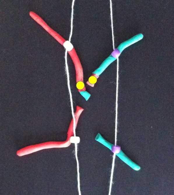
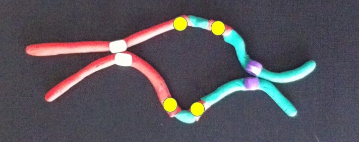
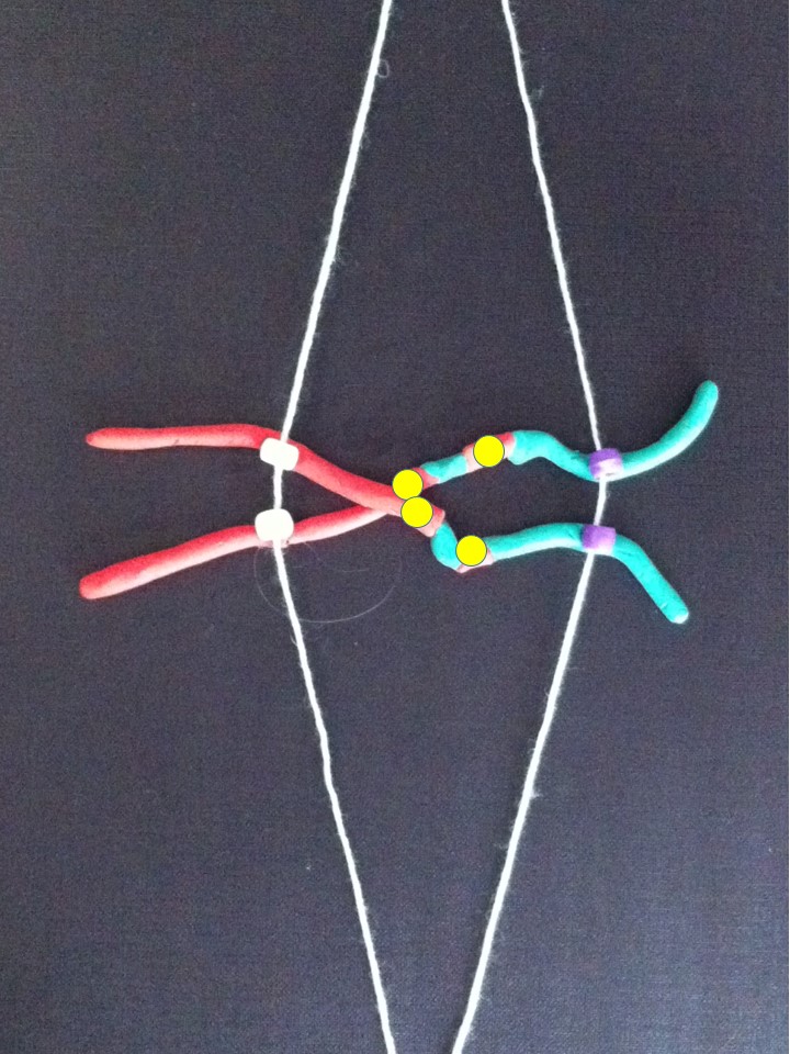
They’re an integral part of our chromosomes. The 46 long strings of genes in each human cell are folded up to form chromosomes, which we can see down the microscope. The telomeres are at both ends of each chromosome. They protect the ends of the chromosomes, and stop the chromosomes from sticking to each other. Chromosomes joining together can be a cause of cancer – more about how that works when we look at the promised breakage-fusion-bridge clay model (in stop motion hopefully!).

The green spots are telomeres on the blue chromosomes from a leukaemia cell. Spot the two abnormal "ring" chromosomes - no ends, no telomeres.

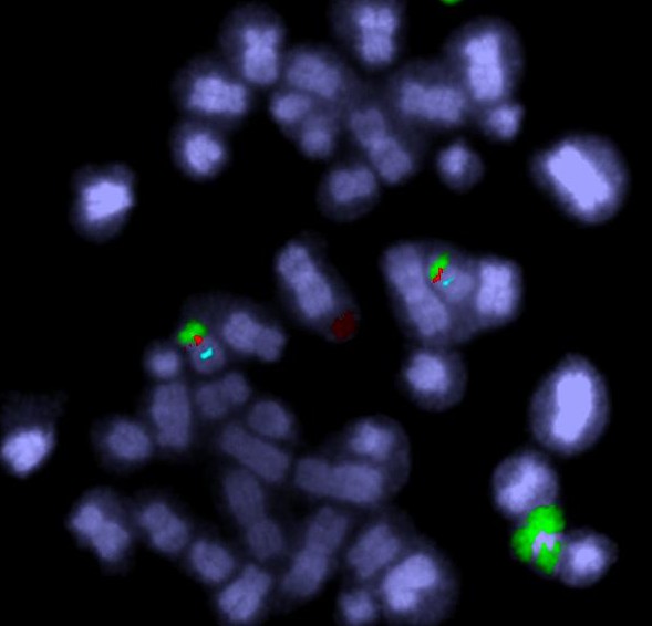
The green spots mark the telomeres on the chromosomes from a leukaemia cell. Spot the two abnormal “ring” chromosomes – no ends, no telomeres (answer next time).

As we age our telomeres get shorter. Telomere shortening has also been associated with other factors such as extreme psychological stress and toxins, including chemotherapy. As well as cancer, short telomeres have been associated with diabetes, cardiovascular disease, osteoarthritis and other diseases. But it also seems that measures like reducing stress, improving diet and exercise may stop or even reverse this premature telomere shortening.

Here’s the paradox – short telomeres can help trigger cancer, but once established the cancer switches on a telomere-lengthening mechanism (usually an enzyme called telomerase) to survive.

And so different researchers are trying contrasting approaches to manipulating telomeres for improved health.

On the one hand, some researchers are looking at the possibility of using the telomere-activating enzyme telomerase to reverse the effects of ageing. Some cosmetics are already available that contain a chemical that’s been reported to activate telomerase. This same chemical is being tested for use as a treatment for some diseases associated with ageing and short telomeres.

On the other hand, because cancers need telomerase to be able to divide indefinitely, other researchers are looking into the possibility of destroying telomerase as a cancer treatment.

These potential treatments will need extensive testing to see if they work and make sure they don’t have unwanted side effects.

From the search for eternal youth to understanding and curing cancer, we haven’t heard the end of telomeres.

INTERESTED IN MORE DETAIL?

A single fertilised egg cell becomes a mature human by growing and dividing into two, many times over. Each time a chromosome makes a copy of itself the telomeres lose a little bit off their ends. The older we get the shorter our telomeres become, so there’s a limited number of times a cell can divide.

That is, unless an enzyme called telomerase is turned on. This enzyme lengthens the telomeres. If you think about it, although most cells in our bodies are programmed to divide a limited number of times, some cells have been dividing for millenia – germ cells – eggs and sperm. It’s cells like these that need telomerase.

Most cancer cells are also able to divide indefinitely by switching on telomerase.

Once the telomeres are dangerously short the chromosomes can start sticking together and becoming abnormal. This stage is called crisis.  The cells don’t function properly, and are a cancer risk, and they stop dividing or self-destruct. There are “tumour suppressors” that look after this self-destruction, but occasionally a cell will bypass this (for example by having a mutated tumour suppressor gene), survive and divide. If the cells divide, these abnormal chromosomes can become more abnormal and turn on cancer genes or lose tumour suppressor genes.

HOW MY RESEARCH FITS IN

This process has mostly been studied in lab animals or cells grown in the laboratory in artificial conditions. So we can extrapolate and suggest that chromosomes with short telomeres can join together and cause cancer. Unfortunately chemotherapy is associated with shortened telomeres, and is a risk factor for leukaemia. These therapy-related leukaemias are usually marked by very abnormal chromosomes.  In my own research I’ve identified some abnormal leukaemia chromosomes that have been made by chromosomes joining together at the telomeres. This was done by identifying which chromosomes are joined together, AND by looking at the molecular content of the chromosomes. End-to-end joining of the chromosomes is actually a lot more common than it’s thought, at least for the abnormal chromosomes I’ve looked at. The similarity between risk factors for very abnormal leukaemia chromosomes and shortened telomeres is interesting.

One thing I’d like to do is find out how common this joining together of the chromosome ends is, in other types of abnormal chromosomes in leukaemia (AML), and eventually look at other cancers. It would help understand how these cancers are caused and possibly identify ways to prevent this. Other information from this type of study could identify more genes with a role in cancer. This opens up new possibilities for developing treatments.

“Abnormal chromosomes made by the end-to-end joining of two chromosomes….” – that sounds like a segue into the breakage-fusion-bridge cycle. More on that later.

FURTHER READING

E. Blackburn and E. Epel 2012. Too toxic to ignore. Nature 490:169-171 (about stress, disease and telomere shortening). Note, Elizabeth Blackburn is Australia’s only female Nobel Prize winner (in science at least) – she shared the prize for Physiology or Medicine in 2009 for her discovery of telomeres.

C. Buseman 2012. Is telomerase a viable target in cancer? Mutation Research 730:90-97

E. Callaway 2010. Telomerase reverses ageing process. Dramatic rejuvenation of prematurely aged mice hints at potential therapy. Nature 28th November 2010 (published online).

B. de Jesus et al. 2013. Telomerase at the intersection of cancer and aging. Trends in Genetics (available online 19th July 2013)

C. Harley et al 2011. A natural product telomerase activator as part of a health maintenance program. Rejuvenation Research 14:45-56.

R. MacKinnon and L. Campbell 2011. The role of dicentric chromosome formation and secondary centromere deletion in the evolution of myeloid malignancy. Genetics Research International Article ID 643628

R. MacKinnon et al 2011. Unbalanced translocation of 20q in AML and MDS often involves interstitial rather than terminal deletion of 20q. Cancer Genetics 204:153-161.

T. Morin. http://www.dayspamagazine.com/article/spa-products-tale-telomeres A balanced article on telomeres in Dayspa Magazine online.

In the year 2000 the draft human genome sequence was announced by Tony Blair and Bill Clinton. It was said to be complete in 2003, in time for the 50th anniversary of the discovery of the structure of DNA. Well actually it wasn’t quite finished. Actually it’s still not finished. Besides the tweaking that still goes on here and there, there are still big gaps. And what’s in these gaps sometimes has a significant role in cancer.

The biggest gaps are centromeres. Centromeres and telomeres are made of what is known as repetitive DNA, and this is hard to sequence. There’s more detail below.

Cancer chromosomes often have centromere or telomere abnormalities. In fact these abnormalities can cause cancer or make it progress faster.

In cancer research there’s a big push to sequence the genomes of different types of cancer  to try and understand the many different DNA changes that can cause cancer. Some researchers try to understand telomeres and centromeres and their role in cancer, but in cancer sequencing projects, and also in diagnostics, centromeres and telomeres are pretty much ignored. Although they’re difficult to sequence, the repetitive DNA does make them easy to study by some other techniques.

One of the goals of personalised medicine is to be able to read a person’s complete genome. For cancer this would include the abnormal cancer genome. But at the moment these gaps mean that we can’t describe the abnormal cancer chromosomes from end to end by sequencing them. The approach I use allows me to work out what’s in each chromosome and discover telomere and centromere abnormalities.

HOW DOES MY RESEARCH FIT IN?

By looking at things that most people don’t worry about I’ve overturned a few assumptions and made some unexpected discoveries, particularly about centromeres in leukaemia.

A normal chromosome has one centromere. I found that chromosomes with two centromeres are more common in  acute myeloid leukaemia (AML) and myelodysplastic syndromes (MDS) than was thought. I found that most of these chromosomes with two centromeres were probably made by two chromosomes joining together.

Telomeres are at the ends of chromosomes and stop them from sticking to each other, so when the telomeres are eroded,  chromosomes can join together. They can be eroded by exposure to chemical toxins, cancer drugs and radiation. So it’s interesting that the leukaemias that are caused by these exposures have more of these two-centromered chromosomes than other leukaemias.

centromeres - dicentric

Fluorescent DNA probes can label up centromeres (blue) and genes (red). In this image there is also a chromosome 20 paint – the green regions are from chromosome 20.

MORE DETAIL ON THE SCIENCE

Telomeres and centromeres are made of highly repetitive DNA and make up some of the gaps in the human genome sequence.

Sequencing a genome is like reading a story. But first we cut the book up into tiny fragments. We read them piece by piece, then try to join the pieces together to make the story, by matching the overlapping parts. Where this approach falls apart is that some sections are repetitive. Some pages are made up of a single word or phrase repeated over and over and over and over and over and over (I won’t repeat that hundreds of thousands of times, but you should get the picture). So if a lot of fragments just say the same thing “over and over and over”, it’s very hard to put them together meaningfully.

The centromere guides the chromosome to the two daughter cells during cell division. A normal chromosome has one centromere. When a chromosome has two centromeres (we call this a dicentric chromosome), the chromosome can be pulled in opposite directions, breaking the chromosome and causing more chromosome disorganisation.Telomeres cap the ends of normal healthy chromosomes. One of their functions is to stop the chromosomes from sticking to each other. So when the telomeres are lost or eroded the chromosomes can join together. That’s one way of creating a chromosome with two centromeres.

Telomere loss is a natural part of ageing. There are also many environmental and lifestyle factors that are thought to affect telomere length. Short telomeres are thought to be a cancer risk because dicentric chromosomes are more likely to arise.

Telomeres and centromeres are very important parts of a normal chromosome. You could say they hold the chromosome together. They have a lot of influence on whether chromosomes are normal and stay normal.

FURTHER READING

Murnane JP 2012. Telomere dysfunction and chromosome instability. Mutat Res. 2012 Feb 1;730(1-2):28-36. (Open access)

MacKinnon RN and Campbell, LJ. 2011. The role of dicentric chromosome formation and secondary centromere deletion in the evolution of myeloid malignancy. Genetics Research International. Article ID 643628. (Open access)

MacKinnon RN, Duivenvoorden HM and Campbell LJ. 2011. Unbalanced translocation of 20q in AML and MDS often involves interstitial rather than terminal deletion of 20q. Cancer Genet. 204(3):153-61.

When it was initiated by the US Department of Energy in 1987, the Human Genome Project was an ambitious, some said impossible, endeavour.  It was all about producing a representative readout of the human genome – that is, the whole set of human DNA.

At the time DNA was read (sequenced) manually – scientists read each letter of the code off an X-ray film. One by one genes were laboriously found and sequenced. Finding and characterising a gene in this way was a whole PhD project, if you got lucky and actually found the gene (finding a gene by family studies was harder than starting with a known protein).

Humans have some 3,000,000,000 letters (base pairs) in their genome, so a faster approach was needed to get the project finished in the planned 15 years.

The Human Genome Project encouraged the development of much faster automated sequencing. I attended the 1991 Cold Spring Harbor Genome Mapping and Sequencing Meeting and there were several examples of exciting new automated sequencing prototypes, which used a range of different approaches.

photo 2

Abstract book from the 1991 Genome Mapping and Sequencing Cold Spring Harbor meeting.
Sample abstracts: “Capillary gel electrophoresis for DNA sequencing – comparison of three different approaches” (HP Swedlow et al); “Library of 256 hexamers, degenerate at two positions (5′-NNXXXX-3′), can create all possible 12-mer primers for applications in high-volulme DNA -sequencing strategies” (D.Shoemaker et al)

Now we can take it for granted that we can look up a gene on the internet. Having a human genome sequence was going to make a big contribution to health care. It is already helping, and will play a bigger role as we learn more about what roles the various genes play. For example working out what genes are playing a role in cancer will become more routine.

We can already do a lot for some cancer patients by doing genetic tests on their cancer. There are many categories of leukaemia that have a very specific type of DNA abnormality, and knowing what gene is involved can help diagnose and treat the disease appropriately.

Chromosome abnormalities helped make some of the earliest cancer gene discoveries. That’s because the gene abnormalities that cause some cancers are caused by microscopically visible changes to the chromosomes, which pinpoint the cancer gene. The poster child for this is chronic myeloid leukaemia. Most cases of CML have a chromosome abnormality known as the Philadelphia translocation. In fact this was the first cancer chromosome abnormality to be discovered. Imatinib (Glivec/Gleevec/STI-571) was one of the first targeted cancer drugs. Designed to lock onto the molecule produced by the cancer gene, it targets the leukaemia cells containing the Philadelphia chromosome. It’s made a huge improvement to the outlook for CML patients.

But for most cancers we’re not so lucky – the cancer-causing genes are not usually so obvious or easy to identify. Most cancers have their own individual combination of genetic errors, and what’s more, the genome changes as the cancer grows more aggressive and spreads. Sequencing of whole cancer genomes could become standard practice in cancer treatment, as a way of understanding each cancer and selecting treatment that targets its specific genetic changes. First we will need to be able to read a complete genome quickly and cheaply. We’re not there yet. But we’re on the way. Compared to 15 years for one representative genome, that’s impressive.

Next time: The Human Genome Project was said to be complete in 2003, in time for the 50th anniversary of the discovery of the structure of DNA. Actually it’s still not finished. Most of the gaps are regions that are very relevant to cancer.

Wellcome_genome_bookcase 2

The first printout of the human genome to be presented as a series of books, displayed in the ‘Medicine Now’ room at the Wellcome Collection, London. The 3.4 billion units of DNA code are transcribed into more than a hundred volumes, each a thousand pages long, in type so small as to be barely legible. From Ross London et al en.wikipedia.

Angelina Jolie decided to have a double mastectomy because she inherited a high risk of breast cancer. What’s remarkable is that all of a sudden people are questioning whether they should be investigating their own cancer risk. Cancer services are fielding many more queries about cancer testing than they were before her announcement. (20th June: I posted this ten days ago, and I’ve just updated the title in response to what has become known as the “Angelina effect”.  Cancer Council Victoria has just announced a 1033% increase in the number of calls to its helpline.)  This is despite the fact that what we know about breast cancer risk that led to Ms Jolie’s decision has already been publicly available for many years. Clearly it was not out there in a way that people took much notice of.

Better communication of science will prevent people from missing or misunderstanding  important information like that.

Is cancer the most common genetic disease? According to The Cancer Genome Project website it is. This might sound scary. Does it mean you have a high risk of cancer if one of your parent has had it? Not necessarily. “Cancer genetics” means one thing to most people and another to cancer scientists. To understand all this we need to understand something about genes and the the role they play.

Genetics is usually understood to mean the characteristics that you inherit from your parents. These characteristics are encoded in our genes,  which are instructions written in a special language called DNA. Every cell has a copy of the set of instructions, and can read them. Genes control our physical features, and the way our bodies’ function, for example how they grow. Genes in the germ cells (eggs and sperm) are passed on from parent to child. Genes in the embryo are passed on to every new cell as we grow. The new cells include eggs and sperm, and their genes are passed on again to the next generation.

The genes can get damaged – this can happen by chance, or it can be caused by environmental factors. Usually this damage is not a problem for a number of reasons. But sometimes the damage is a step in the development of cancer. Usually more than one of these cancerous genetic changes has to occur before cancer is triggered. For breast cancer to develop, these errors need to occur together in the same breast cell.  So cancer can arise in any part of the body, but one of these faulty genes is only inherited if it’s in a germ cell.

If the faulty cancer-promoting genes are inherited, cancer is more likely, because fewer additional gene errors are needed before cancer is triggered. Ms Jolie is reported to have a faulty or mutated BRCA gene. A mutation in a woman’s BRCA gene (BRCA1 or BRCA2) means a 60% lifetime risk of breast cancer, compared to a 12% risk in women without the mutation (National Cancer Institute).

What’s a genome anyway? Scientists studying the genes talk about the genome. This simply means all the genes in an organism – a plant, animal, microbe, you or me. And if you think small-scale, every cell has a genome. Genomics is the study of a genome, be it  the whole organism or a single cell. So “genomics” is really a less ambiguous term than “genetics” when you’re talking about cancer, but to most people it’s jargon. I think this will change as genomics becomes a much more prominent part of health care, including cancer treatment. So to answer the question posed above, yes, cancer is the most common genetic disease, but not in the generally understood sense.

There’s a huge amount of research behind our knowledge of BRCA genes and our ability to use this information to prevent or treat cancer. Cancer is a very diverse disease and we are nowhere near understanding all the gene errors behind all the various types of cancer. The more we discover the better equipped we will be to prevent, detect and cure cancer.

(With thanks to Qian Yu for helpful comments)