Archives for category: genetics

The Leukaemia Foundation of Australia’s National MDS Day has just passed (14th July… but I was busy eating croissants so this post is a little late).

This time I thought I would tell you about a discovery that was made with the help of MDS.

How do healthy cells turn cancerous? Their DNA gradually accumulates errors. Most of these errors aren’t important, but occasionally they stop the cell from working properly. They might cause a cell to grow out of control – and this can lead to cancer.

Myelodysplastic syndromes, or MDS, are a range of blood disorders caused by such errors in the genes. Some types of MDS are relatively mild, but about a third go on to become acute myeloid leukaemia (AML). Thanks to research on MDS we understand its causes a lot better than we did ten or fifteen years ago.

My lab recently published a paper describing three cases of poor prognosis MDS and one case of AML with unusual but remarkably similar changes to the DNA. This complicated structure could not have been predicted by the standard methods of analysing cancer DNA or chromosomes. These features showed us the likely steps that led to these diseases.

Each long string of DNA is folded up neatly to make a chromosome. This is a Claymation that shows how Barbara McClintock’s classic breakage-fusion-bridge cycle causes chromosome abnormalities. The video shows one way that chromosomes (packages of DNA) can become disorganised.

The telomeres (that cap and protect the ends of the chromosomes) are shown falling off, making sticky chromosome ends which join together (see NOTE 2). It’s well accepted that these changes greatly increase the chance of cancerous gene changes. This process has reproduced many, many times in the lab. The problem is that it’s not often been demonstrated in actual cancers. But we did that.
Sometimes only part of the telomere erodes away – enough is lost that it no longer protects the chromosomes from sticking together. But there can be enough telomere DNA left to be a molecular signature of the telomere.

dic 20-22

The arrow points to green dots in the middle of a chromosome. This is the left-over telomere signature that tells us that this abnormal chromosome was made by the joining together of sticky chromosome ends that had their telomeres eroded away. The other green dots are at the chromosome ends. The left and right photos show the same cell but in the right one the abnormal chromosome is identified by its red and blue label.

In our four cases we found that there was a small but non-functional piece of telomere DNA left behind where the two chromosomes joined. Because the telomeres didn’t function, the two chromosome ends could stick together. These caused breakage-fusion-bridge events that caused a protective tumour suppressor gene to be lost, and may have also caused cancer-causing genes to multiply.
MDS and AML have similar genetic causes, so if we learn about the causes of one of them it can help us understand the other. This is often the case with cancer research in a broader sense – if we understand the basic mechanisms in one cancer it can help us understand the mechanisms at work in other cancers better. Telomere fusion could potentially play a role in any cancer, so our MDS research is relevant to cancer research in general.

NOTES

  1. The paper: The dicentric chromosome dic(20;22) is a recurrent abnormality in myelodysplastic syndromes and is a product of telomere fusion. Ruth MacKinnon, Hendrika Duivenvoorden, Lynda Campbell and Meaghan Wall, 2016. Cytogenetic and Genome Research 150(3-4):262-272
  2. The gene errors discussed here usually occur in the body cells rather than the reproductive cells, so they’re not inherited.
  3. For simplicity the Claymation shows telomere fusion in chromosomes that are dividing.  In fact it probably occurs when the DNA is unravelled in the interphase nucleus.
  4. This is cross-posted to Fireside Science on the SciFund Challenge network.

The 14th July is the Leukaemia Foundation of Australia’s annual National MDS Day.

Myelodysplastic Syndromes (MDS) make up a group of diseases that have abnormal blood cell production. MDS is sometimes called pre-leukemia because about a third of patients with MDS will develop leukemia.

MDS is caused by errors in the bone marrow’s genetic information. These errors can often be seen down the microscope as changes to the chromosomes. MDS patients typically have their bone marrow cells analysed to find chromosome abnormalities. Why?

These chromosome abnormalities can reveal important information about their disease, such as diagnosis, appropriate treatment and prognosis.

The IPSS-R is a system that’s used to work out prognosis for MDS patients – that is, how they will do – what their health outlook and risk of developing leukaemia are. A prognostic score is a number calculated from different aspects of the disease. A low score indicates low risk and risk increases as the score goes up. Cytogenetics, or chromosome analysis, is needed to calculate this score because “chromosome abnormalities” is one of the five categories used in the calculation.

For example, if the cells are missing a Y chromosome nothing is added to the IPSS-R prognostic score, whereas if four or more chromosome abnormalities are found, 4 points are added to the score, which can almost single-handedly take the disease into the high (4.5-6) or very high (over 6) risk category.

del20q

Normal chromosome 20 (left) and abnormal chromosome 20 missing most of the long arm (“del(20q)”).


The abnormal chromosome pictured on the right is a deleted chromosome 20  – it’s lost a big chunk carrying hundreds of genes. This is one of the well-known chromosome abnormalities in MDS. We can work out which genes have been lost using higher resolution molecular analysis, but this is not necessary for calculating the IPSS-R prognostic score. One point is added to the score if there’s a deleted chromosome 20 and it’s the only chromosome abnormality. It’s one of the chromosome abnormalities in the “good” cytogenetic category.

So chromosome analysis is an important piece of the puzzle in the care of MDS patients.

More information:

The IPSS-R http://www.bloodjournal.org/content/120/12/2454?sso-checked=true

MDS Foundation – What is MDS? http://www.mds-foundation.org/what-is-mds/

The MDS Beacon http://www.mdsbeacon.com/

Previous MDS Day posts:

Carl Sagan’s Lasts Project – Overcoming MDS

MDS and the Fantastic Mr Dahl

What does IPSS-R stand for? Revised International Prognostic Scoring System for Myelodysplastic Syndromes.

Cross-posted to Fireside Science on the SciFund Challenge network


Most cells in our bodies contain 46 separate long DNA strings that spend most of their time in what appears to be a tangled mess – in a sort of round shape we know as the nucleus. Then lo and behold, these long strings fold up and become chromosomes. Why do they do that?

Bill Earnshaw’s lab at Edinburgh University does some amazing work with chromosomes and cell division. He can explain very elegantly why we need chromosomes.

The DNA makes a copy of itself before the cell divides into two. The chromosomes help make sure each new daughter cell gets an identical copy of this DNA. It’s easier to divide tangled strings into two if you untangle them and roll them up into balls first

Here are some photos from the Earnshaw lab of the chromosomes during cell division.

In the photo above the chromosomes are lining up along the middle of the dividing cell (the “equator” or “metaphase plate”). When they’re all lined up correctly (this stage is “metaphase”) the next stage can start:

The photo above shows the blue chromosome halves (after the doubled-up chromosomes have split in two) separating along the green spindle fibres  (this stage is “anaphase”). Each set of chromosomes will belong to one of the two new daughter cells. If this happens correctly both new cells have identical sets of DNA. This whole cycle of chromosome growth and division is called “mitosis”.

DNA carries genes that make up the blueprint that’s responsible for making every cell, every tissue, every organ work correctly. So it’s important we have the right set of genes.

Cells divide a lot – millions of our cells divide every minute so it’s important that the DNA is shared precisely each time. Mistakes can cause the new daughter cells to misfunction. These cells can become cancerous or produce babies with genetic disease. Usually the cell watches out for these mistakes and self-destructs. But not always. Research helps us understand these processes, how they can go wrong, and work out ways to prevent or fix these mistakes.


Cross-posted to Fireside Science at SciFund Challenge.

Images from http://earnshaw.bio.ed.ac.uk/.

In the 21st century we have an amazing amount of knowledge at our fingertips. This includes the human genome sequence – (most of) the 3,381,944,086 letters of DNA code for a human, known as the human genome sequence. It’s publicly available and you can look it up or download it if you have a computer with internet. Maybe some of us in the developed world are even starting to take it for granted.

The downloadable sequence is just a reference – each one of us has our own unique variation of this standard. Many of us now have reason to find out the sequence of one of our own genes, for example to see if we have inherited a higher than average risk of cancer, or if we carry a gene for a genetic disease. It’s even possible to get your own whole, unique, genome sequenced (at a cost).

In the 1980s the human genome sequence was a dream. Using the technologies available at the time we’d still be sequencing by hand. The Human Genome Project led to a lot of automated sequencing methods being developed – and they’re still being improved, which is still bringing the cost of DNA sequencing down.

This huge project was undertaken by many laboratories across the world. It was intended to be, and has been, a resource for improving health care. It’s not the only sequenced genome. Simpler organisms like viruses and yeasts, which have much smaller genomes, were sequenced before the human genome, and as time goes on we’re getting genome sequences for more and more living things.

So how was the human genome sequenced? There are a range of basic techniques and tools that allow DNA to be manipulated and  read. I’ve put together a Prezi which gives a visual overview of these tools.

Prezi: Tools for DNA discovery and innovation

DNA tools

To see this Prezi with more detailed explanations click on the link above.

DNA can be thought of as a long string of “letters” (nucleotide molecules) strung together. This makes up the code of life and it’s translated into a different language that can make any of the huge variety of proteins, like collagen, haemoglobin, insulin or botox. DNA can be cut, rearranged and joined back together. This makes it very versatile – it can be manipulated. It can be cut up and pasted into different organisms and the host organism will treat it like its own DNA – because the code of life is universal.

Another useful feature of the DNA molecule is that it’s made of two strands which pair together and each one can be made anew from its partner. Thus we can make new DNA in the test tube. One DNA strand can also find its partner so we can find or pick up a whole piece of DNA if we have a  just a small section of its partner.

So these tools for DNA analysis can also be used for our own purposes – for example. to make proteins such as human insulin in massive amounts. You can even make a gene from scratch if you know the code of the protein you want to make. But you’ll still need a live organism to process it into protein for you. Amazing! Diabetics used to rely on pig and cattle insulin, but the human version is better for us!

Cross posted from Fireside Science at SciFund Challenge

Further reading:

The Human Genome Project and cancer

The last frontier of the Human Genome Project – repetitive DNA.

http://www.genome.gov/12011238

The Wellcome Trust’s Sanger Centre has a lot of information, videos and interactive tools that help explain DNA analysis and how the Human Genome was broken down into sections and sequenced. http://www.sanger.ac.uk/about/engagement/yourgenome.html

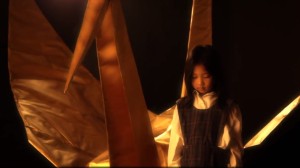
In the summer of 2012-13 my daughter Katherine and her friends got together to make a short film during their holidays while they waited for their University offers.

Nearly two years later here it is.


sadako and golden cranesadako and golden bigsadako and golden boat

Paper Thin is based on the true story of Sadako Sasaki, who tried to fold 1,000 paper cranes to beat her leukaemia. This is an amazing short film directed by Elizabeth Duong with beautiful original music by Daniel Hernandez and Elle Graham. Don’t just take my word for it. Don’t just watch it. Don’t just like it.

Share Paper Thin to help make leukaemia HISTORY.


Mitosis has to be one of the more beautiful things in nature. It’s a choreographed dance of the chromosomes. It’s so small that we can’t see it without a microscope, but it goes on in our bodies billions of times a day.

DNA is a very long molecule made up of the genetic alphabet (which has four letters: A, C, G, T). A gene is made of a certain sequence of DNA letters (or bases) and spells out an instruction for a step in the complex workings of our bodies (such as the structure of insulin). The genes are strung together along the chromosome, and each cell has a set of chromosomes. For our bodies to grow, these cells need to make copies of themselves. The problem of how to distribute the copied chromosomes evenly to the two “daughter cells” is handled very elegantly.


Chromosomes arrested at mitosis and stained with Giemsa (unbanded).

Human metaphase chromosomes stained with Giemsa (unbanded). The two halves of each chromosome are copies of each other.


Mitosis is the solution. Mitosis is broken up into a series of phases: interphase, prophase, metaphase, anaphase, telophase. You could break prophase up further by adding prometaphase: the part of prophase between the nuclear membrane breaking down and metaphase (where the chromosomes line up at the metaphase plate).

Now follow the captions under the pictures.

The interphase nucleus - the DNA from all the chromosomes intertwined with each other is represented by grey modelling clay. (Actually it seems that the chromosomes stay in relatively distinct domains - but under the microscope they appear as one entity.)

The interphase nucleus.   The DNA from all the chromosomes, intermingled with each other, is represented by grey modelling clay. (Actually it seems that the chromosomes stay in relatively distinct domains – but under the microscope they appear as one entity.) The DNA in the interphase nucleus copies itself as the cell grows.


The DNA in the nucleus starts to package and take shape as prophase chromosomes.

The DNA in the nucleus starts to coil up in a pre-determined order and take shape as prophase chromosomes.


The DNA folds up in a pre-determined order to make recognisable chromosomes. When the cell is ready to divide each chromosome has two chromatids or identical halves, joined at the centromere.

The DNA folds up further to make recognisable chromosomes. When the cell is ready to divide each chromosome has two chromatids or identical halves, joined at the centromere.


At metaphase the chromosomes meet in the middle of the cell at the metaphase plate. Then as the cell divides to become two daughter cells, the two halves of the centromere split and travel along the microtubules in opposite directions, pulling the two halves of the chromosome behind them.

Metaphase - the chromosomes line up in the centre of the cell at the metaphase plate. They are attached by their centromeres to microtubules which stretch across the cell.

Metaphase – the chromosomes line up in the centre of the cell at the metaphase plate. They are attached by their centromeres to microtubules which stretch across the cell.


At anaphase the two chromatids (half chromosomes) become the new chromosomes as they separate and move in opposite directions along the microtubules.

At anaphase the two chromatids (half chromosomes) become the new chromosomes as they separate and move in opposite directions along the microtubules.


The chromosomes start to unravel to form the two new daughter interphase nuclei. The cell membrane (the outer covering) pinches at the centre and the one cell finally becomes two (cytokinesis).

The chromosomes start to uncoil to form the two new daughter nuclei – telophase. The cell membrane (the outer covering) pinches at the centre (cytokinesis).


The cell membrane pinches at the centre (cytokinesis) so the cell finally becomes two cells.

Cytokinesis finishes and we have two new cells in interphase.


If a chemical that destroys the microtubules is added to a laboratory culture, the chromosomes are stopped at metaphase. Cytogeneticists (chromosome scientists) use this technique to get enough metaphase chromosomes for analysis. Chromosome banding helps us recognise the chromosomes and identify any changes when an abnormality is suspected. Of course, the cell is also full of other organelles that have to be shared between the new cells.

The modelling clay images above are from my claymation showing mitosis. Modelling clay is a great medium for demonstrating and thinking about how things work, move and change. For the claymation I used a phone camera resting face down on a glass coffee table over the models.

(Cross-posted from Fireside Science at SciFund Challenge.)

Today is Rare Disease Day. Its main aim is to raise awareness among policy makers and the general public. There are over 6,000 rare diseases and 80% of these have a genetic cause.

We have over 20,000 genes coded by over 3,000,000,000 letters of genetic code. Genetic diseases are caused by mistakes in one or more of these genes. These are like typos or spelling mistakes. Not all genetic changes cause disease. But some debilitating genetic diseases are caused by simple one-letter mistakes.

There are also longer mistakes – sections of chromosome can be taken out (deleted) or added in. People with chromosomal syndromes are likely to have several unrelated symptoms, because a cluster of different genes is affected.

The more common genetic diseases are more likely to be researched. There were several labs working to discover the genes for some of the more well-known diseases at the same time – such as cystic fibrosis, Duchenne muscular dystrophy and fragile X syndrome. This competition created a sense of urgency that helped speed up the discovery of these genes. Finding out what gene causes a disease can go a long way to understanding the disease. Sometimes very effective treatments are found by knowing the cause of a disease. The more common genetic diseases that still aren’t fully explained are usually caused by the combined effect of multiple genes. It’s a lot harder to track these down. But there are probably still a lot of rare diseases that could be simply explained but aren’t being researched.

A lack of awareness of rare diseases often means there is misdiagnosis or difficulty getting a diagnosis. People with rare diseases have shared their stories on the Rare Disease Day websites. This is Judit’s story (Judit stars in this video).