Most cells in our bodies contain 46 separate long DNA strings that spend most of their time in what appears to be a tangled mess – in a sort of round shape we know as the nucleus. Then lo and behold, these long strings fold up and become chromosomes. Why do they do that?
Bill Earnshaw’s lab at Edinburgh University does some amazing work with chromosomes and cell division. He can explain very elegantly why we need chromosomes.
The DNA makes a copy of itself before the cell divides into two. The chromosomes help make sure each new daughter cell gets an identical copy of this DNA. It’s easier to divide tangled strings into two if you untangle them and roll them up into balls first
Here are some photos from the Earnshaw lab of the chromosomes during cell division.
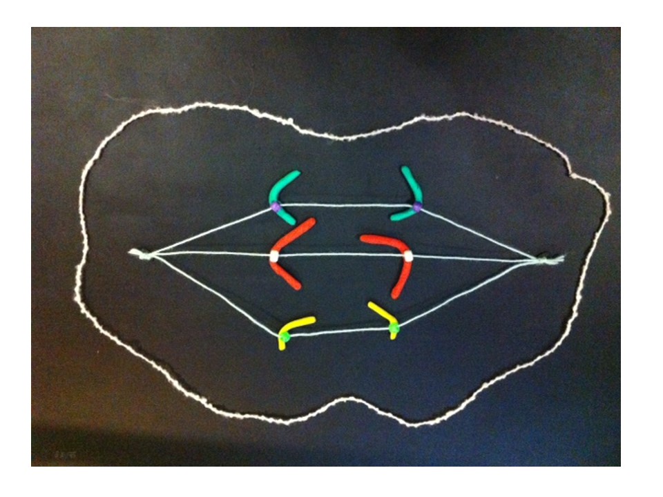
In the photo above the chromosomes are lining up along the middle of the dividing cell (the “equator” or “metaphase plate”). When they’re all lined up correctly (this stage is “metaphase”) the next stage can start:
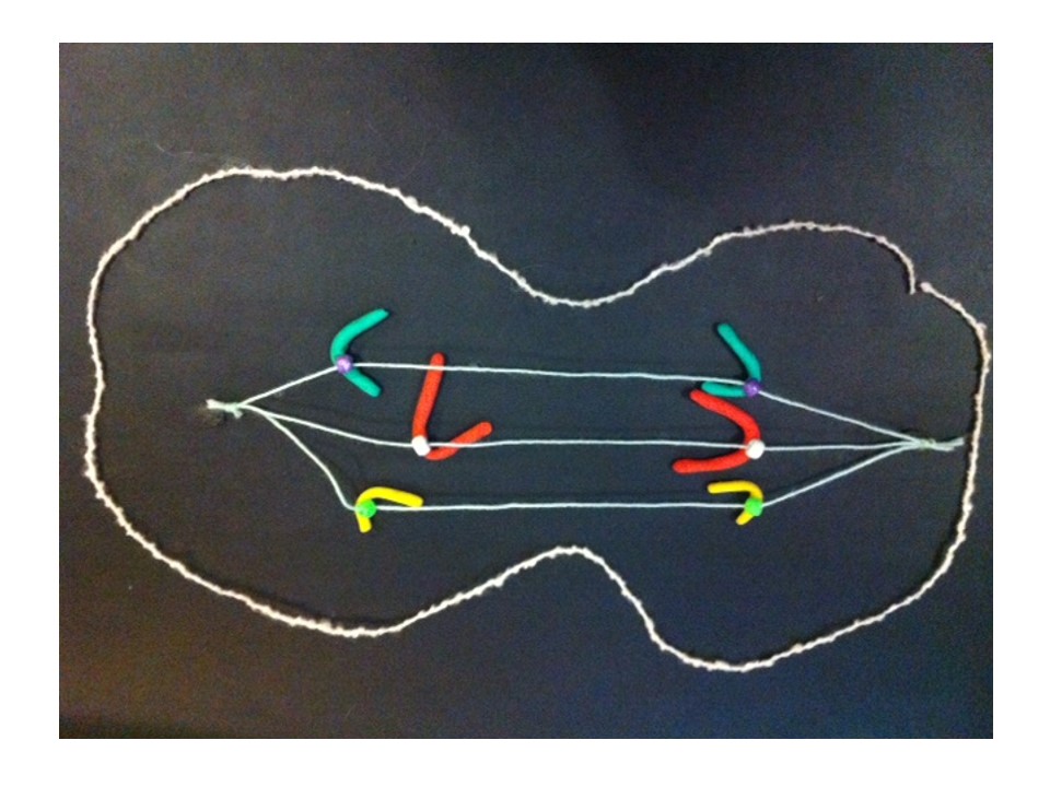
The photo above shows the blue chromosome halves (after the doubled-up chromosomes have split in two) separating along the green spindle fibres (this stage is “anaphase”). Each set of chromosomes will belong to one of the two new daughter cells. If this happens correctly both new cells have identical sets of DNA. This whole cycle of chromosome growth and division is called “mitosis”.
DNA carries genes that make up the blueprint that’s responsible for making every cell, every tissue, every organ work correctly. So it’s important we have the right set of genes.
Cells divide a lot – millions of our cells divide every minute so it’s important that the DNA is shared precisely each time. Mistakes can cause the new daughter cells to misfunction. These cells can become cancerous or produce babies with genetic disease. Usually the cell watches out for these mistakes and self-destructs. But not always. Research helps us understand these processes, how they can go wrong, and work out ways to prevent or fix these mistakes.
Cross-posted to Fireside Science at SciFund Challenge.
Mitosis has to be one of the more beautiful things in nature. It’s a choreographed dance of the chromosomes. It’s so small that we can’t see it without a microscope, but it goes on in our bodies billions of times a day.
DNA is a very long molecule made up of the genetic alphabet (which has four letters: A, C, G, T). A gene is made of a certain sequence of DNA letters (or bases) and spells out an instruction for a step in the complex workings of our bodies (such as the structure of insulin). The genes are strung together along the chromosome, and each cell has a set of chromosomes. For our bodies to grow, these cells need to make copies of themselves. The problem of how to distribute the copied chromosomes evenly to the two “daughter cells” is handled very elegantly.
Human metaphase chromosomes stained with Giemsa (unbanded). The two halves of each chromosome are copies of each other.
Mitosis is the solution. Mitosis is broken up into a series of phases: interphase, prophase, metaphase, anaphase, telophase. You could break prophase up further by adding prometaphase: the part of prophase between the nuclear membrane breaking down and metaphase (where the chromosomes line up at the metaphase plate).
Now follow the captions under the pictures.
The interphase nucleus. The DNA from all the chromosomes, intermingled with each other, is represented by grey modelling clay. (Actually it seems that the chromosomes stay in relatively distinct domains – but under the microscope they appear as one entity.) The DNA in the interphase nucleus copies itself as the cell grows.
The DNA in the nucleus starts to coil up in a pre-determined order and take shape as prophase chromosomes.
The DNA folds up further to make recognisable chromosomes. When the cell is ready to divide each chromosome has two chromatids or identical halves, joined at the centromere.
At metaphase the chromosomes meet in the middle of the cell at the metaphase plate. Then as the cell divides to become two daughter cells, the two halves of the centromere split and travel along the microtubules in opposite directions, pulling the two halves of the chromosome behind them.
Metaphase – the chromosomes line up in the centre of the cell at the metaphase plate. They are attached by their centromeres to microtubules which stretch across the cell.
At anaphase the two chromatids (half chromosomes) become the new chromosomes as they separate and move in opposite directions along the microtubules.
The chromosomes start to uncoil to form the two new daughter nuclei – telophase. The cell membrane (the outer covering) pinches at the centre (cytokinesis).
Cytokinesis finishes and we have two new cells in interphase.
If a chemical that destroys the microtubules is added to a laboratory culture, the chromosomes are stopped at metaphase. Cytogeneticists (chromosome scientists) use this technique to get enough metaphase chromosomes for analysis. Chromosome banding helps us recognise the chromosomes and identify any changes when an abnormality is suspected. Of course, the cell is also full of other organelles that have to be shared between the new cells.
The modelling clay images above are from my claymation showing mitosis. Modelling clay is a great medium for demonstrating and thinking about how things work, move and change. For the claymation I used a phone camera resting face down on a glass coffee table over the models.
Barbara McClintock published a paper describing the breakage-fusion-bridge (BFB) cycle in 1939. Many of her ideas were well before their time. Like many such profound leaps in thinking, the BFB cycle took a long time to catch on. She wrote in 1973, “I stopped publishing detailed reports long ago when I realized, and acutely, the extent of disinterest and lack of confidence in the conclusions I was drawing …One must await the right time for conceptual change.” Her work was appreciated much later and she was awarded a Nobel Prize in 1983 for her discovery of “jumping genes“.
A few weeks back I introduced this post by describing normal chromosome division. This time we’ll look at the breakage-fusion-bridge cycle. This is one way chromosome division can go wrong. Very wrong, in the sense that it can cause the chromosomes to keep changing, and this can cause cancer.
A human chromosome with two centromeres is abnormal. Chromosomes with two centromeres are not unusual in cancer cells. In fact they’re probably a lot more common than we think, because in both research and diagnostic labs the centromeres are usually not looked at.
To recap, a normal chromosome has one centromere. Before the chromosome divides, the two identical halves (chromatids) are held together at the centromere. When the chromosome divides the centromere splits into two halves, the chromatids become the new chromosomes, and the centromeres take the two new chromosomes in different directions into the two new daughter cells.
So what happens if there are two centromeres? If they’re both aligned so that they head in the same direction it’s not a problem – together they take a complete new chromosome with them. The closer the centromeres are together the more likely this is.
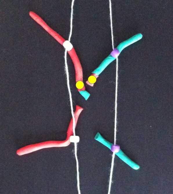
Now follow the pictures and their captions. These describe chromosome division in an abnormal chromosome with two centromeres. Especially follow the yellow dots.
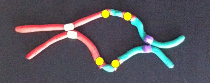
If the two centromeres on a chromosome go in the same direction there’s no problem. But if there’s a twist between the two centromeres when the chromosomes align ready for chromosome division….
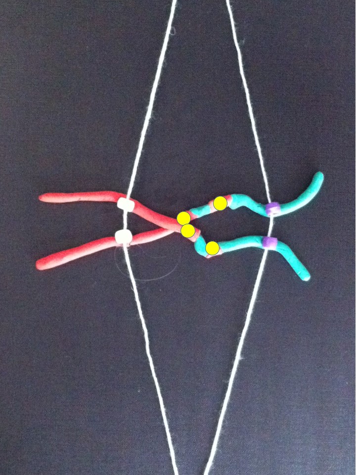
….then, when the two halves of each centromere separate they go in opposite directions. We have a “bridge” spanning the gap between the two centromeres.
The bridge is stretched and can break.
The broken chromosomes in the new cell join together – the top daughter cell gets an extra copy of the yellow gene. The bottom cell loses this copy of the yellow gene.
The new chromosome copies itself to make two equal halves.
If this process repeats..
…
Fusion of the broken pieces creates a chromosome with four copies of the yellow gene.
After replication.
If the yellow gene in the pictures is a cancer gene (“oncogene”) the cell with extra copies might grow and multiply faster than its neighbours. We call this natural selection – the cells that can grow faster than their neighbours become more common which means the genetic change causing that is undergoing “positive selection”. Yes, the cells in our body can evolve and we know this best as cancer.
All this change happens between the two centromeres where the bridge forms. So if we find a chromosome with this type of change on one side of the centromere only it’s a clue that this might have been caused by the breakage-fusion-bridge cycle.
These are modelling clay images from my breakage-fusion-bridge claymation. They’re a bit rough but I hope it helps you understand what happens. Many, perhaps most, images demonstrating the BFB cycle show a different version – where the abnormal chromosome is created by two chromatids of one chromosome breaking and joining together. Most examples don’t show the version I’ve presented – where two different chromosomes have joined together. Check this out on Google Images (search for breakage-fusion-bridge).
Here’s the answer to the quiz from the telomere post. The arrows point to the ring chromosomes. Being rings they have no ends, so no telomeres.
This can happen if the chromosomes are incorrectly distributed to a new cell. The cell could get too many copies of a cancer-promoting gene, or too few copies of a cancer-protecting gene.
The breakage-fusion-bridge cycle is one way that this can happen. This week I was asked to provide a cartoon showing the breakage-fusion-bridge cycle and how it relates to a chromosome abnormality I was describing.
By lucky coincidence my colleague Lan Ta just this week published a paper with a neat breakage-fusion-bridge cartoon that was put together by Bruce Mercer. As well as being a scientist, Bruce is a graphic artist – a very handy combination. So I was trying to draw the modified cartoon for Bruce in two dimensions, without much success. Then modelling clay came to the rescue and I was able to show him what I meant.
So I would like to share the 3D modelling clay version, but before we look at the breakage-fusion-bridge cycle, which is an abnormal pattern of chromosome division, we had better look at normal chromosome division (or mitosis).
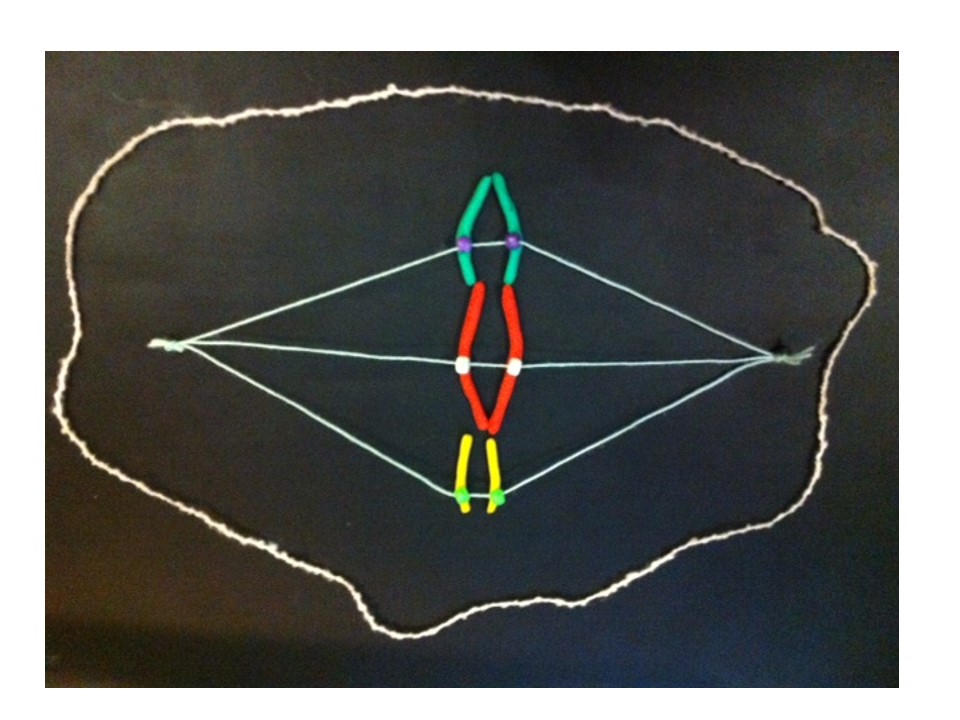
These are chromosomes in modelling clay. There are 23 pairs of chromosomes in a human cell but we will follow 3 chromosomes for simplicity. The chromosomes only take on a recognisable shape when the cell is ready to divide. Each chromosome is made of two halves called chromatids, which are identical. These are held together at the centromere.
In a cell the chromosomes are usually not recognisable – the DNA is unravelled in the nucleus. In a growing cell each unravelled chromosome is producing a copy of itself so that there will be a chromosome for each of the two new daughter cells when the cell divides, or reproduces itself.
After the chromosomes take shape they line up together (at the metaphase plate) between two ends, or poles, of the cell. The centromere has another very important function in cell division. It attaches to fine fibres (microtubules) which stretch between the poles.
The two halves of the centromere separate and each draws its chromatid (which is now a new daughter chromosome) along these microtubules.
So when the cell divides in two to make two new cells, each chromatid becomes a chromosome in one of the new cells.
Cell division complete, the chromosomes unravel and copy themselves again ready for the next cell division.