Archives for posts with tag: Cell Biology

Barbara McClintock published a paper describing the breakage-fusion-bridge (BFB) cycle in 1939. Many of her ideas were well before their time. Like many such profound leaps in thinking, the BFB cycle took a long time to catch on. She wrote in 1973, “I stopped publishing detailed reports long ago when I realized, and acutely, the extent of disinterest and lack of confidence in the conclusions I was drawing …One must await the right time for conceptual change.” Her work was appreciated much later and she was awarded a Nobel Prize in 1983 for her discovery of “jumping genes“.

A few weeks back I introduced this post by describing normal chromosome division. This time we’ll look at the breakage-fusion-bridge cycle. This is one way chromosome division can go wrong. Very wrong, in the sense that it can cause the chromosomes to keep changing, and this can cause cancer.

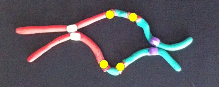
A human chromosome with two centromeres is abnormal. Chromosomes with two centromeres are not unusual in cancer cells. In fact they’re probably a lot more common than we think, because in both research and diagnostic labs the centromeres are usually not looked at.

To recap, a normal chromosome has one centromere. Before the chromosome divides, the two identical halves (chromatids) are held together at the centromere. When the chromosome divides the centromere splits into two halves, the chromatids become the new chromosomes, and the centromeres take the two new chromosomes in different directions into the two new daughter cells.

So what happens if there are two centromeres? If they’re both aligned so that they head in the same direction it’s not a problem – together they take a complete new chromosome with them. The closer the centromeres are together the more likely this is.

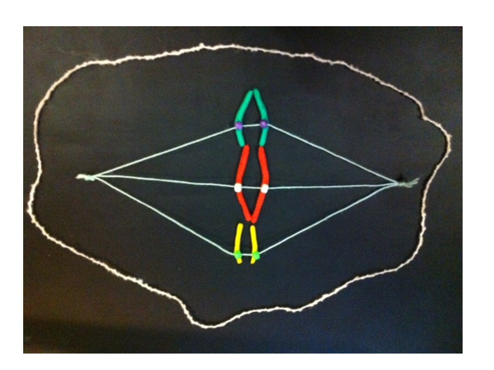
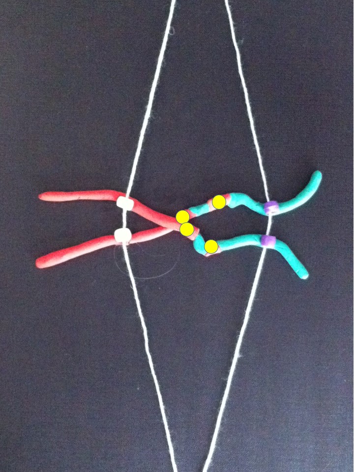
Now follow the pictures and their captions. These describe chromosome division in an abnormal chromosome with two centromeres. Especially follow the yellow dots.

A twist between the two centromeres when the chromosomes align ready for chromosome division.

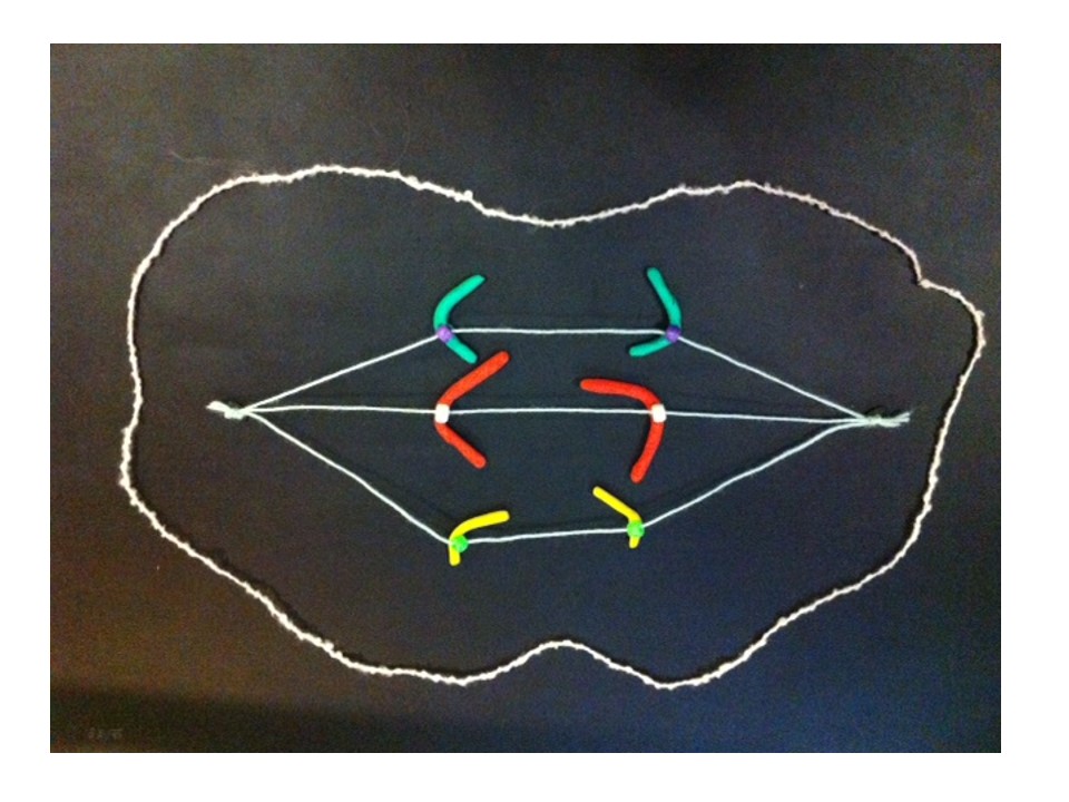
If the two centromeres on a chromosome go in the same direction there’s no problem. But if there’s a twist between the two centromeres when the chromosomes align ready for chromosome division….

....then, when the two halves of each centromere separate they head off in different directions.

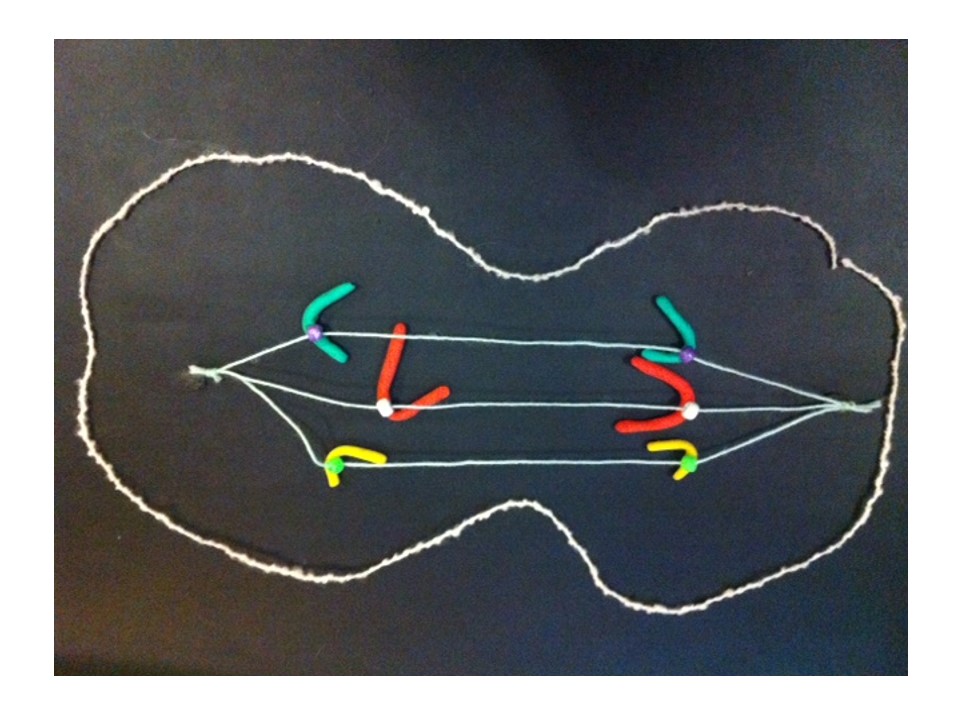
….then, when the two halves of each centromere separate they go in opposite directions. We have a “bridge” spanning the gap between the two centromeres.

The bit of chromosome between them gets stretched and can break.

The bridge is stretched and can break.

The broken chromosomes in the new cell join together - the top daughter cell gets an extra copy of the yellow gene. The bottom cell loses this copy of yellow gene.

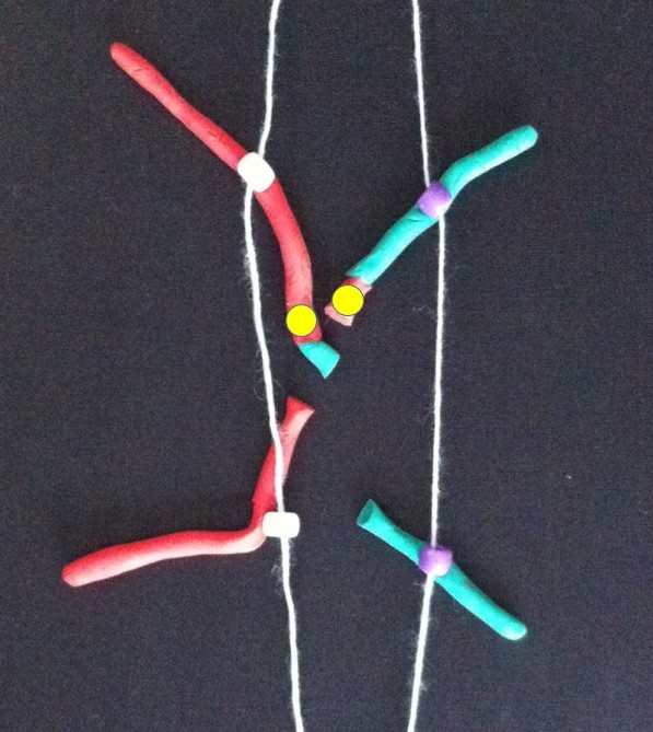
The broken chromosomes in the new cell join together – the top daughter cell gets an extra copy of the yellow gene. The bottom cell loses this copy of the yellow gene.

The new chromosome copies itself to make two equal halves.

The new chromosome copies itself to make two equal halves.

If this process repeats..

If this process repeats..

Fusion of the broken bits of chromosome in the top cell.

Fusion of the broken pieces creates a chromosome with four copies of the yellow gene.

After replication - the new chromosome with four copies of the yellow gene courtesy of the breakage-fusion-bridge cycle.

After replication.

If the yellow gene in the pictures is a cancer gene (“oncogene”) the cell with extra copies might grow and multiply faster than its neighbours. We call this natural selection – the cells that can grow faster than their neighbours become more common which means the genetic change causing that is undergoing “positive selection”. Yes, the cells in our body can evolve and we know this best as cancer.

All this change happens between the two centromeres where the bridge forms. So if we find a chromosome with this type of change on one side of the centromere only it’s a clue that this might have been caused by the breakage-fusion-bridge cycle.

These are modelling clay images from my breakage-fusion-bridge claymation. They’re a bit rough but I hope it helps you understand what happens. Many, perhaps most, images demonstrating the BFB cycle show a different version – where the abnormal chromosome is created by two chromatids of one chromosome breaking and joining together. Most examples don’t show the version I’ve presented – where two different chromosomes have joined together. Check this out on Google Images (search for breakage-fusion-bridge).

Here’s the answer to the quiz from the telomere post. The arrows point to the ring chromosomes. Being rings they have no ends, so no telomeres.

answer to ring chr

Further Reading:

B. McClintock 1939. The Behavior in Successive Nuclear Divisions of a Chromosome Broken at Meiosis. Proc Natl Acad Sci U S A. 1939 August; 25(8): 405–416.

M. Kinsella and V. Bafna 2012. Combinatorics of the Breakage-Fusion-Bridge Mechanism. J Comput Biol. 2012 June; 19(6): 662–678.

R. MacKinnon and L. Campbell 2011. The Role of Dicentric Chromosome Formation and Secondary Centromere Deletion in the Evolution of Myeloid Malignancy. Genetics Research InternationalVolume 2011 (2011), Article ID 643628.

The new Fireside Science blog at SciFund Challenge

I know, it’s been a long time between posts. I’m working on a breakage-fusion-bridge claymation to show how some cancer chromosomes are very changeable. It’s a steep learning curve but hopefully I’ll have it thoroughly mastered soon.

In the meantime here’s a post from Abby Buchwalter in the new group blog “Fireside Science” – SciFund Challenge’s Guide to Life, the Universe, and Everything. Abby and some of the other graduates of the first SciFund Challenge course (including myself) have started a group blog hosted by the SciFund Challenge website. Here we plan to bring the world of Science to readers in an easily digested form.

Abby describes a cell using only the 1,000 most common words in the English language.

Enjoy.

Telomeres. Apparently that’s the new buzz word in cosmetics . They come in different sizes – as we age they shorten. It’s been suggested that we could lengthen them to cancel the effects of ageing, or shorten them to cure cancer. But what are they?

They’re an integral part of our chromosomes. The 46 long strings of genes in each human cell are folded up to form chromosomes, which we can see down the microscope. The telomeres are at both ends of each chromosome. They protect the ends of the chromosomes, and stop the chromosomes from sticking to each other. Chromosomes joining together can be a cause of cancer – more about how that works when we look at the promised breakage-fusion-bridge clay model (in stop motion hopefully!).

The green spots are telomeres on the blue chromosomes from a leukaemia cell. Spot the two abnormal "ring" chromosomes - no ends, no telomeres.

The green spots mark the telomeres on the chromosomes from a leukaemia cell. Spot the two abnormal “ring” chromosomes – no ends, no telomeres (answer next time).

As we age our telomeres get shorter. Telomere shortening has also been associated with other factors such as extreme psychological stress and toxins, including chemotherapy. As well as cancer, short telomeres have been associated with diabetes, cardiovascular disease, osteoarthritis and other diseases. But it also seems that measures like reducing stress, improving diet and exercise may stop or even reverse this premature telomere shortening.

Here’s the paradox – short telomeres can help trigger cancer, but once established the cancer switches on a telomere-lengthening mechanism (usually an enzyme called telomerase) to survive.

And so different researchers are trying contrasting approaches to manipulating telomeres for improved health.

On the one hand, some researchers are looking at the possibility of using the telomere-activating enzyme telomerase to reverse the effects of ageing. Some cosmetics are already available that contain a chemical that’s been reported to activate telomerase. This same chemical is being tested for use as a treatment for some diseases associated with ageing and short telomeres.

On the other hand, because cancers need telomerase to be able to divide indefinitely, other researchers are looking into the possibility of destroying telomerase as a cancer treatment.

These potential treatments will need extensive testing to see if they work and make sure they don’t have unwanted side effects.

From the search for eternal youth to understanding and curing cancer, we haven’t heard the end of telomeres.

INTERESTED IN MORE DETAIL?

A single fertilised egg cell becomes a mature human by growing and dividing into two, many times over. Each time a chromosome makes a copy of itself the telomeres lose a little bit off their ends. The older we get the shorter our telomeres become, so there’s a limited number of times a cell can divide.

That is, unless an enzyme called telomerase is turned on. This enzyme lengthens the telomeres. If you think about it, although most cells in our bodies are programmed to divide a limited number of times, some cells have been dividing for millenia – germ cells – eggs and sperm. It’s cells like these that need telomerase.

Most cancer cells are also able to divide indefinitely by switching on telomerase.

Once the telomeres are dangerously short the chromosomes can start sticking together and becoming abnormal. This stage is called crisis.  The cells don’t function properly, and are a cancer risk, and they stop dividing or self-destruct. There are “tumour suppressors” that look after this self-destruction, but occasionally a cell will bypass this (for example by having a mutated tumour suppressor gene), survive and divide. If the cells divide, these abnormal chromosomes can become more abnormal and turn on cancer genes or lose tumour suppressor genes.

HOW MY RESEARCH FITS IN

This process has mostly been studied in lab animals or cells grown in the laboratory in artificial conditions. So we can extrapolate and suggest that chromosomes with short telomeres can join together and cause cancer. Unfortunately chemotherapy is associated with shortened telomeres, and is a risk factor for leukaemia. These therapy-related leukaemias are usually marked by very abnormal chromosomes.  In my own research I’ve identified some abnormal leukaemia chromosomes that have been made by chromosomes joining together at the telomeres. This was done by identifying which chromosomes are joined together, AND by looking at the molecular content of the chromosomes. End-to-end joining of the chromosomes is actually a lot more common than it’s thought, at least for the abnormal chromosomes I’ve looked at. The similarity between risk factors for very abnormal leukaemia chromosomes and shortened telomeres is interesting.

One thing I’d like to do is find out how common this joining together of the chromosome ends is, in other types of abnormal chromosomes in leukaemia (AML), and eventually look at other cancers. It would help understand how these cancers are caused and possibly identify ways to prevent this. Other information from this type of study could identify more genes with a role in cancer. This opens up new possibilities for developing treatments.

“Abnormal chromosomes made by the end-to-end joining of two chromosomes….” – that sounds like a segue into the breakage-fusion-bridge cycle. More on that later.

FURTHER READING

E. Blackburn and E. Epel 2012. Too toxic to ignore. Nature 490:169-171 (about stress, disease and telomere shortening). Note, Elizabeth Blackburn is Australia’s only female Nobel Prize winner (in science at least) – she shared the prize for Physiology or Medicine in 2009 for her discovery of telomeres.

C. Buseman 2012. Is telomerase a viable target in cancer? Mutation Research 730:90-97

E. Callaway 2010. Telomerase reverses ageing process. Dramatic rejuvenation of prematurely aged mice hints at potential therapy. Nature 28th November 2010 (published online).

B. de Jesus et al. 2013. Telomerase at the intersection of cancer and aging. Trends in Genetics (available online 19th July 2013)

C. Harley et al 2011. A natural product telomerase activator as part of a health maintenance program. Rejuvenation Research 14:45-56.

R. MacKinnon and L. Campbell 2011. The role of dicentric chromosome formation and secondary centromere deletion in the evolution of myeloid malignancy. Genetics Research International Article ID 643628

R. MacKinnon et al 2011. Unbalanced translocation of 20q in AML and MDS often involves interstitial rather than terminal deletion of 20q. Cancer Genetics 204:153-161.

T. Morin. http://www.dayspamagazine.com/article/spa-products-tale-telomeres A balanced article on telomeres in Dayspa Magazine online.

What makes a cell become cancerous?

This can happen if the chromosomes are incorrectly distributed to a new cell. The cell could get too many copies of a cancer-promoting gene, or too few copies of a cancer-protecting gene.

The breakage-fusion-bridge cycle is one way that this can happen. This  week I was asked to provide a cartoon showing the breakage-fusion-bridge cycle and how it relates to a chromosome abnormality I was describing.

By lucky coincidence my colleague Lan Ta just this week published a paper with a neat breakage-fusion-bridge cartoon that was put together by Bruce Mercer. As well as being a scientist, Bruce is a graphic artist – a very handy combination. So I was trying to draw the modified cartoon for Bruce in two dimensions, without much success. Then modelling clay came to the rescue and I was able to show him what I meant.

So I would like to share the 3D modelling clay version, but before we look at the breakage-fusion-bridge cycle, which is an abnormal pattern of chromosome division, we had better look at normal chromosome division (or mitosis).

image

These are chromosomes in modelling clay. There are 23 pairs of chromosomes in a human cell but we will follow 3 chromosomes for simplicity.  The chromosomes only take on a recognisable shape when the cell is ready to divide. Each chromosome is made of two halves called chromatids, which are identical. These are held together at the centromere.

mitosis 1

In a cell the chromosomes are usually not recognisable – the DNA is unravelled in the nucleus. In a growing cell each unravelled  chromosome is producing a copy of itself so that there will be a chromosome for each of the two new daughter cells when the cell divides, or reproduces itself.

Slide3

After the chromosomes take shape they line up together (at the metaphase plate) between two ends, or poles, of the cell. The centromere has another very important function in cell division. It attaches to fine fibres (microtubules) which stretch between the poles.

Slide4

Slide5

The two halves of the centromere separate and each draws its chromatid (which is now a new daughter chromosome) along these microtubules.

Slide6

Slide7

Slide8

So when the cell divides in two to make two new cells, each chromatid becomes a chromosome in one of the new cells.

Slide9

Cell division complete, the chromosomes unravel and copy themselves again ready for the next cell division.

This process happens successfully millions of times every day to create new cells in our bodies – amazing.