What makes a cell become cancerous?
This can happen if the chromosomes are incorrectly distributed to a new cell. The cell could get too many copies of a cancer-promoting gene, or too few copies of a cancer-protecting gene.
The breakage-fusion-bridge cycle is one way that this can happen. This week I was asked to provide a cartoon showing the breakage-fusion-bridge cycle and how it relates to a chromosome abnormality I was describing.
By lucky coincidence my colleague Lan Ta just this week published a paper with a neat breakage-fusion-bridge cartoon that was put together by Bruce Mercer. As well as being a scientist, Bruce is a graphic artist – a very handy combination. So I was trying to draw the modified cartoon for Bruce in two dimensions, without much success. Then modelling clay came to the rescue and I was able to show him what I meant.
So I would like to share the 3D modelling clay version, but before we look at the breakage-fusion-bridge cycle, which is an abnormal pattern of chromosome division, we had better look at normal chromosome division (or mitosis).

These are chromosomes in modelling clay. There are 23 pairs of chromosomes in a human cell but we will follow 3 chromosomes for simplicity. The chromosomes only take on a recognisable shape when the cell is ready to divide. Each chromosome is made of two halves called chromatids, which are identical. These are held together at the centromere.

In a cell the chromosomes are usually not recognisable – the DNA is unravelled in the nucleus. In a growing cell each unravelled chromosome is producing a copy of itself so that there will be a chromosome for each of the two new daughter cells when the cell divides, or reproduces itself.

After the chromosomes take shape they line up together (at the metaphase plate) between two ends, or poles, of the cell. The centromere has another very important function in cell division. It attaches to fine fibres (microtubules) which stretch between the poles.
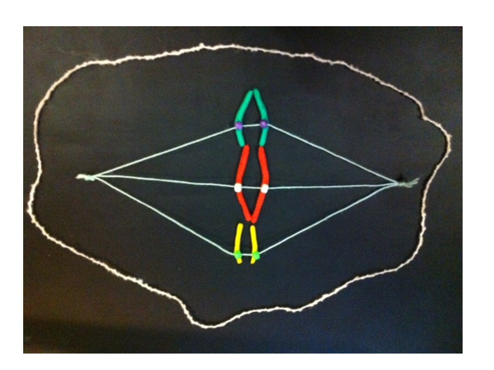

The two halves of the centromere separate and each draws its chromatid (which is now a new daughter chromosome) along these microtubules.
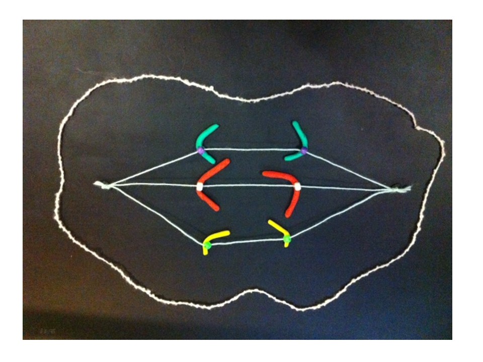
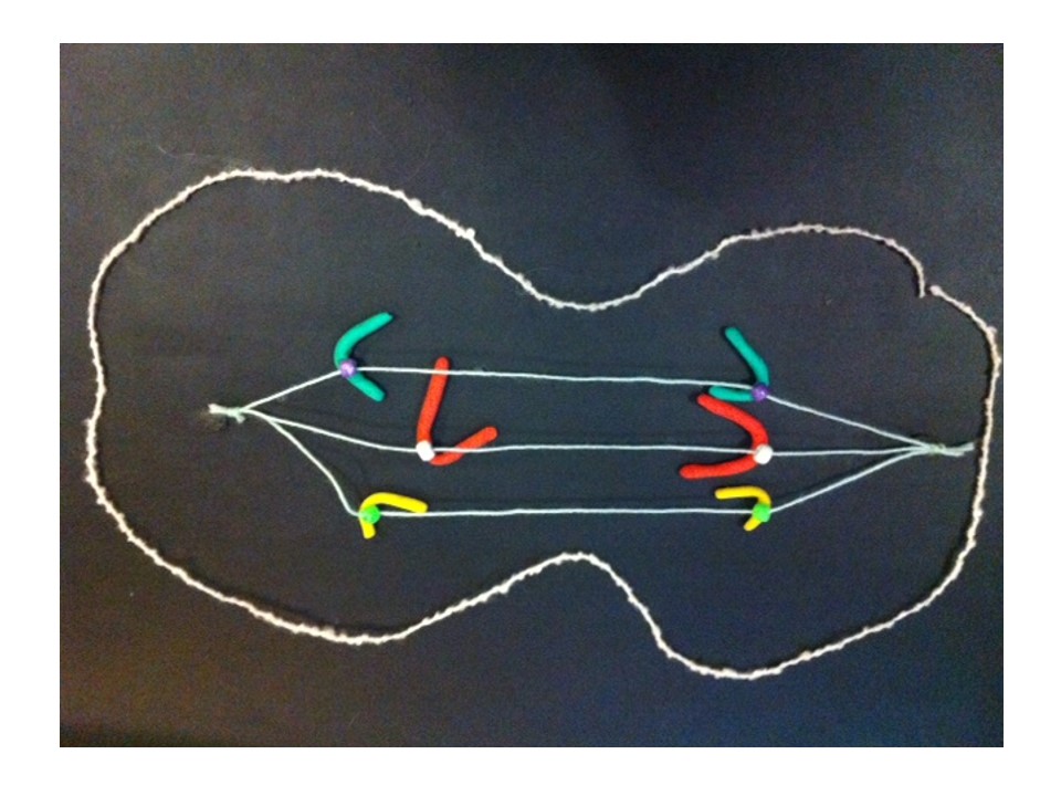

So when the cell divides in two to make two new cells, each chromatid becomes a chromosome in one of the new cells.

Cell division complete, the chromosomes unravel and copy themselves again ready for the next cell division.
This process happens successfully millions of times every day to create new cells in our bodies – amazing.

[…] few weeks back I introduced this post by describing normal chromosome division. This time we’ll look at the […]