Archives for category: chromosomes

The Leukaemia Foundation of Australia’s National MDS Day has just passed (14th July… but I was busy eating croissants so this post is a little late).

This time I thought I would tell you about a discovery that was made with the help of MDS.

How do healthy cells turn cancerous? Their DNA gradually accumulates errors. Most of these errors aren’t important, but occasionally they stop the cell from working properly. They might cause a cell to grow out of control – and this can lead to cancer.

Myelodysplastic syndromes, or MDS, are a range of blood disorders caused by such errors in the genes. Some types of MDS are relatively mild, but about a third go on to become acute myeloid leukaemia (AML). Thanks to research on MDS we understand its causes a lot better than we did ten or fifteen years ago.

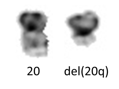
My lab recently published a paper describing three cases of poor prognosis MDS and one case of AML with unusual but remarkably similar changes to the DNA. This complicated structure could not have been predicted by the standard methods of analysing cancer DNA or chromosomes. These features showed us the likely steps that led to these diseases.

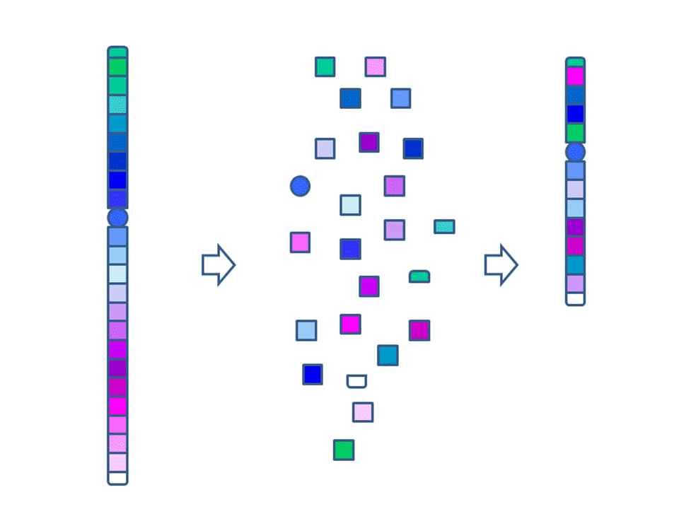
Each long string of DNA is folded up neatly to make a chromosome. This is a Claymation that shows how Barbara McClintock’s classic breakage-fusion-bridge cycle causes chromosome abnormalities. The video shows one way that chromosomes (packages of DNA) can become disorganised.

The telomeres (that cap and protect the ends of the chromosomes) are shown falling off, making sticky chromosome ends which join together (see NOTE 2). It’s well accepted that these changes greatly increase the chance of cancerous gene changes. This process has reproduced many, many times in the lab. The problem is that it’s not often been demonstrated in actual cancers. But we did that.
Sometimes only part of the telomere erodes away – enough is lost that it no longer protects the chromosomes from sticking together. But there can be enough telomere DNA left to be a molecular signature of the telomere.

dic 20-22

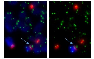
The arrow points to green dots in the middle of a chromosome. This is the left-over telomere signature that tells us that this abnormal chromosome was made by the joining together of sticky chromosome ends that had their telomeres eroded away. The other green dots are at the chromosome ends. The left and right photos show the same cell but in the right one the abnormal chromosome is identified by its red and blue label.

In our four cases we found that there was a small but non-functional piece of telomere DNA left behind where the two chromosomes joined. Because the telomeres didn’t function, the two chromosome ends could stick together. These caused breakage-fusion-bridge events that caused a protective tumour suppressor gene to be lost, and may have also caused cancer-causing genes to multiply.
MDS and AML have similar genetic causes, so if we learn about the causes of one of them it can help us understand the other. This is often the case with cancer research in a broader sense – if we understand the basic mechanisms in one cancer it can help us understand the mechanisms at work in other cancers better. Telomere fusion could potentially play a role in any cancer, so our MDS research is relevant to cancer research in general.

NOTES

  1. The paper: The dicentric chromosome dic(20;22) is a recurrent abnormality in myelodysplastic syndromes and is a product of telomere fusion. Ruth MacKinnon, Hendrika Duivenvoorden, Lynda Campbell and Meaghan Wall, 2016. Cytogenetic and Genome Research 150(3-4):262-272
  2. The gene errors discussed here usually occur in the body cells rather than the reproductive cells, so they’re not inherited.
  3. For simplicity the Claymation shows telomere fusion in chromosomes that are dividing.  In fact it probably occurs when the DNA is unravelled in the interphase nucleus.
  4. This is cross-posted to Fireside Science on the SciFund Challenge network.

In the cells that make up your body, about 2 metres (6 feet) of DNA – strings of genes – are coiled up and packaged into a typically roundish nucleus. This nucleus is only about one hundred-thousandth of a metre wide. I’ve said before that the DNA packed into the nucleus “appears to be a tangled mess”. But looks can be deceptive.

In this great little video Carl Zimmer challenges the idea that DNA in the nucleus is arranged randomly. Job Dekker and his group are finding that the nucleus is actually very organised. Zimmer conjures up an image of tiny robots in the cell folding the DNA very precisely.

//players.brightcove.net/245991542/344c319b-6d23-4cbc-975e-c8530534af8a_default/index.html?videoId=4823709456001

[From Science Happens! Episode 5: Everything you thought you knew about the shape of DNA is wrong]

Some genes control other genes. Dekker’s research suggests that the way DNA folds helps this process – by bringing controlling genes close to the genes they regulate. One cause of cancer could be bad folding that interferes with this control mechanism.

Dekker’s group is trying to work out which genes lie next to each other and how DNA is folded. The answer will be extremely complex.  Dekker thinks that one day this knowledge will make it possible to fix badly folded DNA in cancer cells.

Cross-posted to Fireside Science on the SciFund Challenge network.

The 14th July is the Leukaemia Foundation of Australia’s annual National MDS Day.

Myelodysplastic Syndromes (MDS) make up a group of diseases that have abnormal blood cell production. MDS is sometimes called pre-leukemia because about a third of patients with MDS will develop leukemia.

MDS is caused by errors in the bone marrow’s genetic information. These errors can often be seen down the microscope as changes to the chromosomes. MDS patients typically have their bone marrow cells analysed to find chromosome abnormalities. Why?

These chromosome abnormalities can reveal important information about their disease, such as diagnosis, appropriate treatment and prognosis.

The IPSS-R is a system that’s used to work out prognosis for MDS patients – that is, how they will do – what their health outlook and risk of developing leukaemia are. A prognostic score is a number calculated from different aspects of the disease. A low score indicates low risk and risk increases as the score goes up. Cytogenetics, or chromosome analysis, is needed to calculate this score because “chromosome abnormalities” is one of the five categories used in the calculation.

For example, if the cells are missing a Y chromosome nothing is added to the IPSS-R prognostic score, whereas if four or more chromosome abnormalities are found, 4 points are added to the score, which can almost single-handedly take the disease into the high (4.5-6) or very high (over 6) risk category.

del20q

Normal chromosome 20 (left) and abnormal chromosome 20 missing most of the long arm (“del(20q)”).


The abnormal chromosome pictured on the right is a deleted chromosome 20  – it’s lost a big chunk carrying hundreds of genes. This is one of the well-known chromosome abnormalities in MDS. We can work out which genes have been lost using higher resolution molecular analysis, but this is not necessary for calculating the IPSS-R prognostic score. One point is added to the score if there’s a deleted chromosome 20 and it’s the only chromosome abnormality. It’s one of the chromosome abnormalities in the “good” cytogenetic category.

So chromosome analysis is an important piece of the puzzle in the care of MDS patients.

More information:

The IPSS-R http://www.bloodjournal.org/content/120/12/2454?sso-checked=true

MDS Foundation – What is MDS? http://www.mds-foundation.org/what-is-mds/

The MDS Beacon http://www.mdsbeacon.com/

Previous MDS Day posts:

Carl Sagan’s Lasts Project – Overcoming MDS

MDS and the Fantastic Mr Dahl

What does IPSS-R stand for? Revised International Prognostic Scoring System for Myelodysplastic Syndromes.

Cross-posted to Fireside Science on the SciFund Challenge network


Chromosome-folding theory shows promise | EurekAlert! Science News.

Most cells in our bodies contain 46 separate long DNA strings that spend most of their time in what appears to be a tangled mess – in a sort of round shape we know as the nucleus. Then lo and behold, these long strings fold up and become chromosomes. Why do they do that?

Bill Earnshaw’s lab at Edinburgh University does some amazing work with chromosomes and cell division. He can explain very elegantly why we need chromosomes.

The DNA makes a copy of itself before the cell divides into two. The chromosomes help make sure each new daughter cell gets an identical copy of this DNA. It’s easier to divide tangled strings into two if you untangle them and roll them up into balls first

Here are some photos from the Earnshaw lab of the chromosomes during cell division.

In the photo above the chromosomes are lining up along the middle of the dividing cell (the “equator” or “metaphase plate”). When they’re all lined up correctly (this stage is “metaphase”) the next stage can start:

The photo above shows the blue chromosome halves (after the doubled-up chromosomes have split in two) separating along the green spindle fibres  (this stage is “anaphase”). Each set of chromosomes will belong to one of the two new daughter cells. If this happens correctly both new cells have identical sets of DNA. This whole cycle of chromosome growth and division is called “mitosis”.

DNA carries genes that make up the blueprint that’s responsible for making every cell, every tissue, every organ work correctly. So it’s important we have the right set of genes.

Cells divide a lot – millions of our cells divide every minute so it’s important that the DNA is shared precisely each time. Mistakes can cause the new daughter cells to misfunction. These cells can become cancerous or produce babies with genetic disease. Usually the cell watches out for these mistakes and self-destructs. But not always. Research helps us understand these processes, how they can go wrong, and work out ways to prevent or fix these mistakes.


Cross-posted to Fireside Science at SciFund Challenge.

Images from http://earnshaw.bio.ed.ac.uk/.

As we’ve seen in previous posts, cancer is caused by some sort of error in the DNA of the cancer. Human DNA comes in 46 long strings called chromosomes and it sometimes breaks, but luckily the break is usually repaired. However, sometimes the repair process gets it wrong – for example two DNA ends are joined together that aren’t meant to be together.

This post is about a fairly recent discovery called chromothripsis. I’m going to describe it in terms of language, because after all, DNA is a language, and I think it will help picture what’s going on.

Anagrams are made by rearranging all the letters of a phrase or word to make another phrase or word, and it’s best if the new has a similar meaning to the old. Some examples are:

astronomer —-> moon starer
the meaning of life —-> the fine game of nil.

The rule for “perfect” anagrams is that all the letters are re-used in the new sentence.

Setting up printing originally involved arranging individual letters in a particular order on a plate. If the letters were dropped it wouldn’t be easy to get the letters back in the right order. If you had to put them back together in a hurry they’d be jumbled up and you wouldn’t get a meaningful anagram. In fact you’d probably have some missing letters left on the floor in a corner somewhere. Ok it’s starting to get complicated, but something similar can happen to DNA!

A printer inspecting a large form of type on a cylinder press. Each of the islands of text represents a single page, the darker blocks are images. The whole bed of type is printed on a single sheet of paper, which is then folded and cut to form many individual pages of a book.

At the beginning of 2011 Joshua Stephens and his colleagues published a paper that got a lot of cancer biologists excited. They noticed that the DNA of some cancers was very messed up. They even said that they’d found this in 10% of the cancers where they looked for it. Not only that, but they thought this had happened all at once by shattering of the chromosomes and rejoining of the broken pieces in a random and totally incorrect order.

Scientists love to make complicated words out of Greek roots. The Stephens paper called this process, this shattering and stitching back together of the chromosomes, “chromothripsis“. (An aside: The chromo- in chromothripsis is short for chromosome, or coloured thing. The authors say that thripsis means shattering into pieces.)

I had a couple of nice examples of chromothripsis acute myeloid leukaemia, but as someone who’s interested in chromosomes I wanted to talk about the chromosomes that were made by this whole chromothripsis process. There was no word for these new chromosomes so I came up with the term “anachromosome“. I like it because it has connotations of “new”, “remade”, and my favourite, “anagrams”. Although the anagram rule says all the letters have to be reused, there are apparently imperfect” anagrams which can leave out some letters to make the anagram work. This is more like an anachromosome. Inevitably lots of bits of chromosome don’t make it into the anachromosome and so are lost. Such wholesale shuffling and loss of large sections of DNA will probably kill the cell in most cases, but just occasionally it will produce a cell that can outgrow its neighbours – and turn cancerous.

This shows how the chromosome shatters and rejoins in a random order. Note that the two ends of the chromosome and the centromere (represented by a circle) are preserved. These are essential features of a functioning linear chromosome. From MacKinnon and Campbell 2013. Cancer Genetics 206:238-251.

This shows how the chromosome shatters and rejoins in a random order. Note that the two ends of the chromosome and the centromere (represented by a circle) are preserved. These are essential features of a functioning linear chromosome. From MacKinnon and Campbell 2013. Cancer Genetics 206:238-251.

References:

MacKinnon RN and Campbell LJ 2013. Chromothripsis under the microscope: a cytogenetic perspective of two cases of AML with catastrophic chromosome rearrangement. Cancer Genetics 206:238-251

Stephens PJ et al. 2011. Massive Genomic Rearrangement Acquired in a Single Catastrophic Event during Cancer Development. Cell 144:27-40


Carl Sagan was an astronomer and academic, best known for popularising astronomy. He hosted and co-produced the original hugely popular series Cosmos: A Personal Voyage.  Its sequel Cosmos: A Spacetime Odyssey was released this year. Even though I’m a biologist at heart I was fascinated by the original Cosmos.

Sagan was diagnosed with a myelodysplastic syndrome (MDS) and died at the age of 62, in 1996. In interviews near the end of his life he discussed myelodysplasia and said he was hopeful he’d been cured. He died at the Fred Hutchinson Cancer Research Center of pneumonia  after his third bone marrow transplant, a complication of this illness.

Most people with a diagnosis of MDS won’t have heard of it before. MDS is a group of bone marrow diseases. It’s at least as common as or more common than leukaemia but older people have a higher risk – perhaps one in 2,000 over the age of 60. A third of people with MDS will develop leukaemia. The 14th July, 2014, is the Leukaemia Foundation of Australia‘s second National MDS Day . One of the aims of MDS Day is to raise awareness of MDS.

Sagan’s illness was an opportunity to popularise MDS, but look how the cause of his death was described in these TV news reports.

In these news stories he was said to have died from a complication of “a rare blood disorder that led to cancer”, or “a blood disease”, “a bone marrow disease”, and even a” bone cancer” – the name of his disease was avoided.

Myelodysplasia literally means abnormal bone marrow cells. Blood cells are made in the bone marrow. In MDS the immature bone marow cells are abnormal and don’t mature properly. So the blood doesn’t have enough normal blood cells to do its job effectively. The blood is made of a number of different types of cells and the different types of MDS relate to the type of abnormal cell. MDS is often associated with a recognised chromosome abnormality, and identifying these chromosome abnormalities can help with diagnosis, treatment and prognosis. Therapy-related MDS is a specific type of MDS caused by treatment for a previous unrelated cancer and it usually has a poor outcome and very abnormal chromosomes.

MDS research has been neglected but has picked up recently. Some of the recent progress includes work by Carl Walkley and Louise Purton at St Vincent’s Institute in Melbourne, Australia.

MDS has had a history of name changes that seems to have made the meaning of its name less clear, except to medically trained people. This hasn’t helped improve public awareness of MDS. It was first named Di Guglielmo Syndrome in 1923 after its discoverer, then became refractory anaemia, then preleukaemic anaemia, preleukaemic acute human leukaemia, preleukaemia, and finally in 1976 the French-American-British Co-Operative Group of haematologists named it myelodysplastic syndromes. This recognised that it’s a group of related diseases and that not all cases will go on to develop into leukaemia.

Pathologist Ed Uthman, thinks Sagan’s Disease would be a better name for myelodysplastic syndromes – both as a tribute to Carl Sagan and a name that would mean more to most people than myelodysplastic syndromes.  Maybe he has something. Plenty of syndromes and diseases are named after people who studied them. Down Syndrome would have to be the best known example. Have you heard of amyotrophic lateral sclerosis? Motor neurone disease? Lou Gehrig’s disease? The first name is probably a nice technical description of the disease, but I’m guessing you’re more likely to  have an idea of what the disease is from one of the last two names, because they’re used in popular media and are connected in the public eye with famous sufferers – Stephen Hawking and Lou Gehrig. (Ed Uthman also think’s Lou Gehrig’s Disease should be “Hawking’s Disease”.)

I’ll let Carl Sagan have the last words on popular (mis)understanding of science (extract from Wikiquote).

We live in a society absolutely dependent on science and technology and yet have cleverly arranged things so that almost no one understands science and technology. That’s a clear prescription for disaster.

Every kid starts out as a natural-born scientist, and then we beat it out of them. A few trickle through the system with their wonder and enthusiasm for science intact.

(Cross-posted to Fireside Science at SciFund Challenge.)

Mitosis has to be one of the more beautiful things in nature. It’s a choreographed dance of the chromosomes. It’s so small that we can’t see it without a microscope, but it goes on in our bodies billions of times a day.

DNA is a very long molecule made up of the genetic alphabet (which has four letters: A, C, G, T). A gene is made of a certain sequence of DNA letters (or bases) and spells out an instruction for a step in the complex workings of our bodies (such as the structure of insulin). The genes are strung together along the chromosome, and each cell has a set of chromosomes. For our bodies to grow, these cells need to make copies of themselves. The problem of how to distribute the copied chromosomes evenly to the two “daughter cells” is handled very elegantly.


Chromosomes arrested at mitosis and stained with Giemsa (unbanded).

Human metaphase chromosomes stained with Giemsa (unbanded). The two halves of each chromosome are copies of each other.


Mitosis is the solution. Mitosis is broken up into a series of phases: interphase, prophase, metaphase, anaphase, telophase. You could break prophase up further by adding prometaphase: the part of prophase between the nuclear membrane breaking down and metaphase (where the chromosomes line up at the metaphase plate).

Now follow the captions under the pictures.

The interphase nucleus - the DNA from all the chromosomes intertwined with each other is represented by grey modelling clay. (Actually it seems that the chromosomes stay in relatively distinct domains - but under the microscope they appear as one entity.)

The interphase nucleus.   The DNA from all the chromosomes, intermingled with each other, is represented by grey modelling clay. (Actually it seems that the chromosomes stay in relatively distinct domains – but under the microscope they appear as one entity.) The DNA in the interphase nucleus copies itself as the cell grows.


The DNA in the nucleus starts to package and take shape as prophase chromosomes.

The DNA in the nucleus starts to coil up in a pre-determined order and take shape as prophase chromosomes.


The DNA folds up in a pre-determined order to make recognisable chromosomes. When the cell is ready to divide each chromosome has two chromatids or identical halves, joined at the centromere.

The DNA folds up further to make recognisable chromosomes. When the cell is ready to divide each chromosome has two chromatids or identical halves, joined at the centromere.


At metaphase the chromosomes meet in the middle of the cell at the metaphase plate. Then as the cell divides to become two daughter cells, the two halves of the centromere split and travel along the microtubules in opposite directions, pulling the two halves of the chromosome behind them.

Metaphase - the chromosomes line up in the centre of the cell at the metaphase plate. They are attached by their centromeres to microtubules which stretch across the cell.

Metaphase – the chromosomes line up in the centre of the cell at the metaphase plate. They are attached by their centromeres to microtubules which stretch across the cell.


At anaphase the two chromatids (half chromosomes) become the new chromosomes as they separate and move in opposite directions along the microtubules.

At anaphase the two chromatids (half chromosomes) become the new chromosomes as they separate and move in opposite directions along the microtubules.


The chromosomes start to unravel to form the two new daughter interphase nuclei. The cell membrane (the outer covering) pinches at the centre and the one cell finally becomes two (cytokinesis).

The chromosomes start to uncoil to form the two new daughter nuclei – telophase. The cell membrane (the outer covering) pinches at the centre (cytokinesis).


The cell membrane pinches at the centre (cytokinesis) so the cell finally becomes two cells.

Cytokinesis finishes and we have two new cells in interphase.


If a chemical that destroys the microtubules is added to a laboratory culture, the chromosomes are stopped at metaphase. Cytogeneticists (chromosome scientists) use this technique to get enough metaphase chromosomes for analysis. Chromosome banding helps us recognise the chromosomes and identify any changes when an abnormality is suspected. Of course, the cell is also full of other organelles that have to be shared between the new cells.

The modelling clay images above are from my claymation showing mitosis. Modelling clay is a great medium for demonstrating and thinking about how things work, move and change. For the claymation I used a phone camera resting face down on a glass coffee table over the models.

(Cross-posted from Fireside Science at SciFund Challenge.)

Cancer has been described as the most common genetic disease. This doesn’t necessarily mean it’s hereditary – usually the genetic mistakes that cause cancer arise in the body’s organs or tissues and can’t be inherited. We’re continually learning of new cancer-causing genetic mistakes.

If we think of genes as words spelling out the instructions for our bodies to function, there are different types of mistakes or “typos” that can cause cancer. Some of these are like spelling mistakes – an incorrect letter or two. The mistakes I’ll be talking about here involve whole words (or lots of them). For example one or more copies of a word are added – ” very big” becomes “very very big” – extra copies of a cancer gene (we call them oncogenes) can cause or accelerate cancer growth. Or if a word is lost – “don’t grow” becomes “grow” – this illustrates loss of a tumour suppressor gene.

I described the breakage-fusion-bridge cycle a few weeks back. The BFB cycle was a theory developed from studies with maize, but it also applies to some cancers. This is an example of basic research,  inspired by curiosity but eventually being useful in ways we never imagined. If you look back at that post it shows how the BFB cycle can cause gain or loss of genes. If a cancer gene is  multiplied, or if a tumour suppressor gene is in the part that’s lost, the cell can gain a growth advantage over other cells, which is part of the process causing cancer.

Here’s an example.

Cancer cell lines are cancer cells that can be grown indefinitely in the laboratory. I’ve just published a paper on HEL, which is a leukaemia cell line. It’s popular for studying how cells make globin (molecules in red blood cells that help us process the air we breathe).

The chromosomes of HEL are very abnormal. I’ve used a combination of techniques to show how the chromosomes are reorganised as well as which parts have been lost, gained and amplified.  It’s very complicated.

One of the gene abnormalities in HEL is amplification of the JAK2 gene. JAK2 is a well-known cancer gene that is often abnormal in blood cancers. The normal gene can be mutated to become a cancer gene, for example by a “spelling mistake” in the DNA. By adding extra copies of this abnormal gene the effects can be magnified. This is known as gene amplification. There are a few cancer genes that are commonly amplified in cancers.

To cut a long story short, JAK2 is amplified in the HEL cell line. And a nearby tumour suppressor gene (CDKN2A) has been lost. But only by looking at the chromosomes does the reason become clear. Some detective work tells us that there were some breakage-fusion-bridge events. I won’t go into the detail – if you’re interested it’s in the paper. But we have chromosomes whose ancestors had two centromeres, and if we use a DNA tag for the region between the centromeres we can see “stripes”.

Here’s an example from HEL that shows DNA amplified by the BFB cycle – we can show where a gene is on the chromosomes by labelling it with a fluorescence tagged DNA “probe”. The striped pattern reminds us of the yellow dots in the modelling clay demonstration:

The red is DNA that's normally at one end of some of the chromosomes. The stripes tell us that the end of a chromosome (22) is in the middle of these chromosomes and there are extra copies. This helps us work out how these chromosomes were made. It's a strong clue that BFB cycles were involved and the ancestral chromosome had two centromeres.

The red is DNA that’s normally at one end of some of the chromosomes. The stripes tell us that the end of a chromosome (22) is in the middle of these chromosomes and there are extra copies. It’s a strong clue that BFB cycles were involved and the ancestral chromosome had two centromeres.

The new chromosome with four copies of the yellow gene courtesy of the breakage-fusion-bridge cycle.

The new chromosome with four copies of the yellow gene courtesy of the breakage-fusion-bridge cycle.

Recently JAK2 amplification was also reported in triple-negative breast cancer. Triple negative means that three well-known genetic causes of breast cancer are not present. So finding JAK2 amplification would help explain the cause of some triple-negative breast cancers, and could help work out an effective treatment. Perhaps this JAK2 amplification is sometimes caused by BFB cycles. Without looking at the layout of the abnormal chromosomes we may never know.

To end this story, here’s Bruce Mercer’s cartoon diagram showing the BFB cycle in HEL: http://emph.oxfordjournals.org/content/2013/1/225/F6.expansion.html

Chromosomes that cause cancer. Part 2. The Philadelphia Chromosome.

This is a link to my latest post on the Fireside Science Blog on the SciFund Challenge website.

It describes the first discovery of a cancer-causing chromosome and the exciting progress in treating chronic myeloid leukaemia that it made possible.